Image of Rhabditophora
Description:
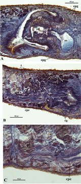
Figure 5.Dugesia bifida. Microphotographs of the copulatory apparatus. A Holotype ZMA V.Pl. 7189.1, sagittal section showing the penis bulb (pb) and the penis papilla (pp) with the seminal vesicle (sv) and the ejaculatory duct (ed) B Holotype ZMA V.Pl. 7189.1, sagittal section showing the opening of the right oviducal branch (rob) through the posterior wall of the bursal canal (bc), and the common posterior oviducal extension (cpe) full of sperm (s) C Paratype CGAS Pla 7.1, sagittal section showing the caudal part of the common posterior oviducal extension (cpe) and the ductule (du) communicating with the ventral part of an adjacent vitellarium (v).
Included On The Following Pages:
- Eukaryota (eukaryotes)
- Opisthokonta (opisthokonts)
- Metazoa (Animal)
- Bilateria
- Protostomia (protostomes)
- Platyhelminthes (flatworms)
- Rhabditophora
- Life (creatures)
- Cellular (cellular organisms)
- Spiralia (spiralians)
- Tricladida (planarians)
- Geoplanoidea
- Dugesiidae
- Dugesia
- Dugesia bifida
This image is not featured in any collections.
Source Information
- license
- cc-by-3.0
- copyright
- Giacinta Angela Stocchino, Ronald Sluys, Renata Manconi
- bibliographic citation
- Stocchino G, Sluys R, Manconi R (2014) A new and aberrant species of Dugesia (Platyhelminthes, Tricladida, Dugesiidae) from Madagascar ZooKeys 425: 71–88
- original
- original media file
- visit source
- partner site
- Zookeys
- ID