Image of Neomonachus Slater & Helgen
Description:
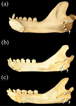
Figure 11.Medial view of right dentaries of a Monachus monachus b Neomonachus schauinslandi, and c Neomonachus tropicalis. The mandibular foramen is situated inferior to the mandibular notch in Monachus, and opens immediately to the medial surface of the ramus. In Neomonachus, the foramen is anteriorly displaced and is set in a groove or sulcus that extends from inferior to the mandibular notch. Also note the expanded rugose area for insertion of the pterygoid muscles in Monachus. This region is poorly developed in Neomonachus.
Included On The Following Pages:
This image is not featured in any collections.
Source Information
- license
- cc-by-3.0
- copyright
- Dirk-Martin Scheel, Graham J. Slater, Sergios-Orestis Kolokotronis, Charles W. Potter, David S. Rotstein, Kyriakos Tsangaras, Alex D. Greenwood, Kristofer M. Helgen
- bibliographic citation
- Scheel D, Slater G, Kolokotronis S, Potter C, Rotstein D, Tsangaras K, Greenwood A, Helgen K (2014) Biogeography and taxonomy of extinct and endangered monk seals illuminated by ancient DNA and skull morphology ZooKeys 409: 1–33
- original
- original media file
- visit source
- partner site
- Zookeys
- ID