Image of Copa

Description:
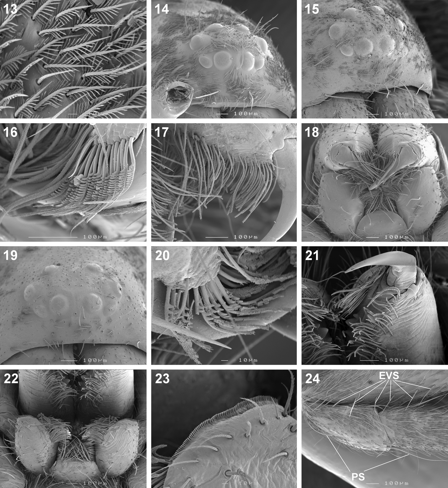
Figures 13–24.Scanning electron microscope photographs of Copa flavoplumosa Simon, 1885 female (13, 14, 16) and male (15, 17, 18) and Copa kei sp. n. female (19–24): 13 dorsal carapace setae 14, 15, 19 eye region and clypeus, anterolateral (14, 15) and anterior (19) views 16, 17, 20 cheliceral promarginal bent setae, anterior view 18, 22 mouthparts, ventral view 21 chelicerae, ventral view 23 serrula 24 femur, patella and tibia of leg II, indicating erect ventral setae on femora (EVS) and proximal and distal dorsal patellar setae (PS).
Included On The Following Pages:
- Life (creatures)
- Cellular (cellular organisms)
- Eukaryota (eukaryotes)
- Opisthokonta (opisthokonts)
- Metazoa (Animal)
- Bilateria
- Protostomia (protostomes)
- Ecdysozoa (ecdysozoans)
- Arthropoda (arthropods)
- Chelicerata (chelicerates)
- Arachnida (arachnids)
- Araneae (spiders)
- Opisthothelae
- Araneomorphae
- Entelegynae
- Retrolateral tibial apophysis
- Corinnidae (antmimics and ground sac spiders)
- Copa
- Copa flavoplumosa
- Panarthropoda
This image is not featured in any collections.
Source Information
- license
- cc-by-3.0
- copyright
- Charles Richard Haddad
- bibliographic citation
- Haddad C (2013) A revision of the continental species of Copa Simon, 1885 (Araneae, Corinnidae) in the Afrotropical Region ZooKeys 276: 1–37
- original
- original media file
- visit source
- partner site
- Zookeys
- ID