Image of Selenops
Description:
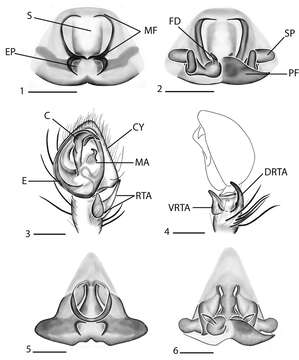
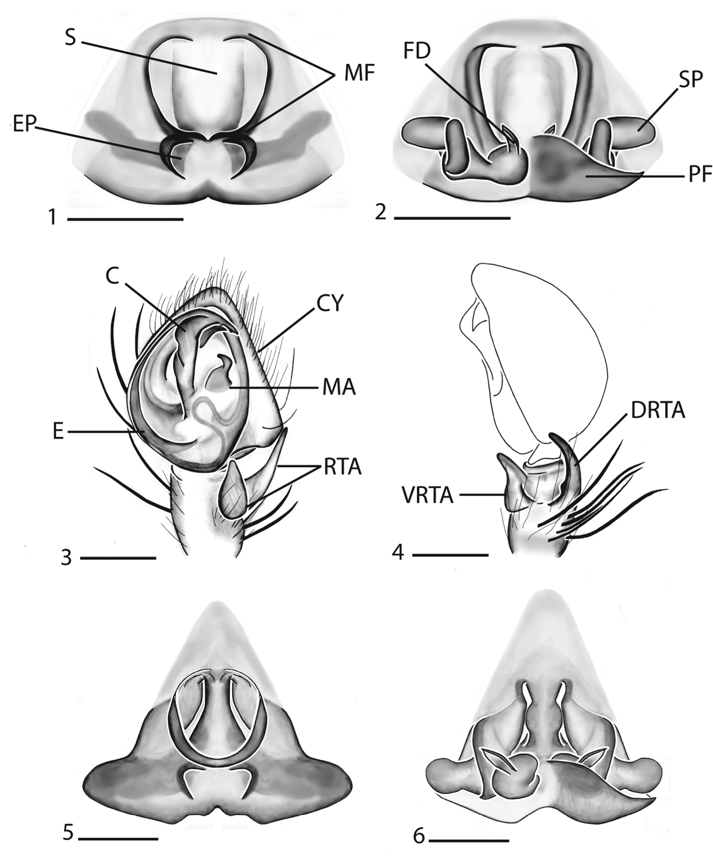
Figures 1–6.Copulatory organs of Selenops arikok sp. n. female holotype from Arikok National Park, Aruba (EME sel_068) 1–2 Selenops curazao Alayón-García male holotype from CarMaBI, Curaçao, Netherlands Antilles (MCZ) 3–4 female paratype from Piscadera Baai building, Curaçao, Netherlands Antilles (MCZ) 5–6, 1, 5 epigyne, ventral view 2, 6 spermathecae, dorsal view 3 male pedipalp, ventral view 4 male pedipalp, retrolateral view. Scale bar = 0.40 mm (1–2), 0.30 mm (3–6). Abbreviations: S = septum, MF = median field, EP = epigynal pockets, FD = fertilization duct, SP = spermathecae, PF = posterodorsal fold, C = conductor, CY = cymbium, MA = median apophysis, E = embolus, RTA = retrolateral tibial apophysis, VRTA = ventral retrolateral tibial apophysis, DRTA = dorsal retrolateral tibial apophysis.
Included On The Following Pages:
- Life (creatures)
- Cellular (cellular organisms)
- Eukaryota (eukaryotes)
- Opisthokonta (opisthokonts)
- Metazoa (Animal)
- Bilateria
- Protostomia (protostomes)
- Ecdysozoa (ecdysozoans)
- Arthropoda (arthropods)
- Chelicerata (chelicerates)
- Arachnida (arachnids)
- Araneae (spiders)
- Opisthothelae
- Araneomorphae
- Entelegynae
- Retrolateral tibial apophysis
- Selenopidae (wall crab spiders)
- Selenops
- Selenops arikok
- Panarthropoda
This image is not featured in any collections.
Source Information
- license
- cc-by-3.0
- copyright
- Sarah C. Crews
- bibliographic citation
- Crews S (2011) A revision of the spider genus Selenops Latreille, 1819 (Arachnida, Araneae, Selenopidae) in North America, Central America and the Caribbean ZooKeys 105: 1–182
- original
- original media file
- visit source
- partner site
- Zookeys
- ID