black and white portrait
Description:
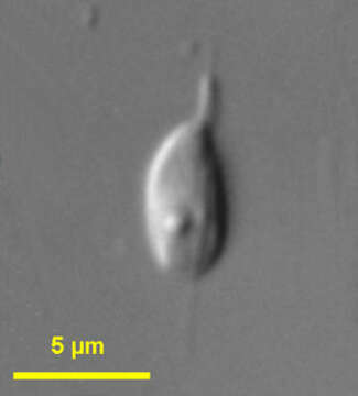
Amastigomonas (a-ma-stig-owe-moan-ass) debruynei de Saedeleer, 1931. Cells are 5 to 6.5 microns long, dorso-ventrally flattened and flexible but not amoeboid. The anterior flagellum emerges from the tip of a laterally directed sleeve and beats in a small angle. The posterior flagellum is slightly longer than the length of the cell, lies in a groove along the margin of the cell, trails under the cell and occasionally protrudes behind the cell. Strands of cytoplasm may be drawn out behind the cell. The nucleus is situated in the anterior left of the cell. Commonly observed.
Included On The Following Pages:
- Life (creatures)
- Cellular (cellular organisms)
- Eukaryota (eukaryotes)
- Apusomonadidae (apusomonads)
- Amastigomonas
- Amastigomonas debruynei
This image is not featured in any collections.
Source Information
- license
- cc-by-nc
- author
- Won Je Lee
- provider
- micro*scope
- original
- original media file
- visit source
- partner site
- micro*scope
- ID