Surface detail
Description:
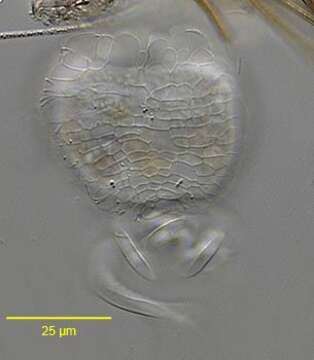
Surface detail of the peritrich ciliate, Pseudovorticella chlamydophora (Penard,1922) Jankowski, 1976. This genus is distinguished from the genus Vorticella by its grid-like silver line system. The transverse components of the silverline system of Vorticella species have no vertical connections. P. Chlamydophora has a thick clear pellicular layer composed of cuboid units, which give the cell surface a distinctive quilted appearance (seen en face here). The extended cell is an inverted bell shape connected at the aboral scopula to a contractile stalk. The cell is spherical when contracted. The stalk contracts as a coil rather than a zigzag (e.g. Haplocaulus). The otherwise identical P. vestita has two contractile vacuoles. P. chlamydophora may be gregarious but does not form true colonies. Collected from a freshwater pond near Boise, Idaho.June 2005. DIC.
Included On The Following Pages:
- Life (creatures)
- Cellular (cellular organisms)
- Eukaryota (eukaryotes)
- SAR (Stramenopiles, Alveolates, Rhizaria)
- Alveolata (alveolates)
- Ciliophora (ciliates)
- Intramacronucleata
- Oligohymenophorea
- Peritrichia
- Sessilida
- Vorticellidae
- Pseudovorticella
- Pseudovorticella chlamydophora
This image is not featured in any collections.
Source Information
- license
- cc-by-nc
- author
- William Bourland
- provider
- micro*scope
- original
- original media file
- visit source
- partner site
- micro*scope
- ID