portrait
Description:
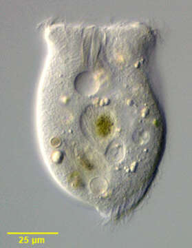
Portrait of the Phyllopharyngeid ciliate, Phascolodon vorticella (Stein, 1859). The genus is probably monotypic. P. vorticella is easily recognized by its distinctive horse-saddle shape. The cell is dorsoventrally compressed with a deep ventral furrow that bears 5-6 kineties on the right and 8-10 kineties on the left. There is one transverse file of cilia on the dorsal surface (i.e. the dorsal brush). The right kineties curve around the flared anterior end. The posterior tapers to a blunt point. At the anterior end of the ventral furrow is a slit-like cytostome with a prominent dorsally directed cytopharyngeal basket of nematodesmata. The coarsely granular macronucleus is located in the posterior ½. There are two contractile vacuoles, one on the right anterior side of the ventral furrow and the other on the left posterior side of it. P. vorticella is planktonic and feeds mainly on algae and cyanobacteria. Collected from freshwater pond near Boise, Idaho December 2004. DIC optics.
Included On The Following Pages:
- Life (creatures)
- Cellular (cellular organisms)
- Eukaryota (eukaryotes)
- SAR (Stramenopiles, Alveolates, Rhizaria)
- Alveolata (alveolates)
- Ciliophora (ciliates)
- Intramacronucleata
- Phyllopharyngea
- Phyllopharyngia
- Chlamydodontida
- Chilodonellidae
- Phascolodon
- Phascolodon vorticella
This image is not featured in any collections.
Source Information
- license
- cc-by-nc
- author
- William Bourland
- provider
- micro*scope
- original
- original media file
- visit source
- partner site
- micro*scope
- ID