oral
Description:
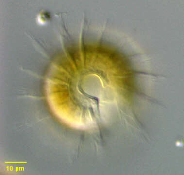
Portrait (anterior apical view) of the oligotrich ciliate, Pelagostrombidium mirabile (Penard, 1916; Krainer 1991). This genus was established (Krainer, 1991) for strombidiids differing from other Strombidium species in having an interrupted equatorial extrusome girdle, and a globular neoformation organelle (site of formation of daughter cell oral apparatus). The similar freshwater genus, Limnostrombidium differs from Pelagostrombidium in having a much smaller differently oriented cytostome and uninterrupted extrusome girdle and tubular neoformation organelle. P. mirabile differs from P. fallax in that the latter is reddish due to numerous tiny cytoplasmic pigment granules. The cytoplasm of both P. fallax and P. mirabile contains sequestered chloroplasts (i.e. kleptoplasts) from ingested algae. The similar freshwater genus, Limnostrombidium differs from Pelagostrombidium in having a much smaller differently oriented cytostome and uninterrupted extrusome girdle and tubular neoformation organelle. The shape of P. mirabile is obovoid with a projecting collar anteriorly (C-shaped in this en face view). A yellow-green color is imparted by sequestered functional plastids (i.e. kleptoplasts). There is a prominent open wreath of adoral membranelles (seen well here). The cytostome (seen at four o'clock position in this image) is ~1/2 the body length. An equatorial girdle of ~15 ? long extrusomes is interrupted by the cytostome. The globular macronucleus is central. A layer of polygonal polysaccharide plates covers most of the cell surface. The neoformation organelle is seen as a striated structure in the posterior 1/3 in this image. P. mirabile is a rapid swimmer. Collected from freshwater agricultural irrigation canal near Boise, Idaho October 2003. DIC optics.
Included On The Following Pages:
- Life (creatures)
- Cellular (cellular organisms)
- Eukaryota (eukaryotes)
- SAR (Stramenopiles, Alveolates, Rhizaria)
- Alveolata (alveolates)
- Ciliophora (ciliates)
- Intramacronucleata
- Spirotrichea
- Oligotrichia
- Strombidiidae
- Pelagostrombidium
- Pelagostrombidium mirabile
This image is not featured in any collections.
Source Information
- license
- cc-by-nc
- author
- William Bourland
- provider
- micro*scope
- original
- original media file
- visit source
- partner site
- micro*scope
- ID