Drawing
Description:
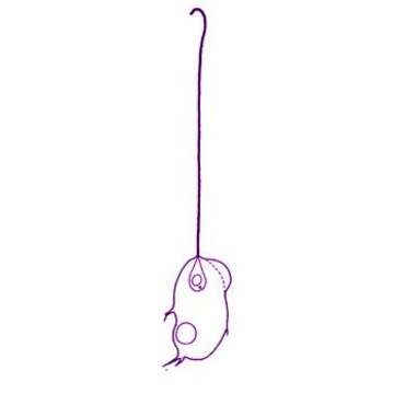
Mastigamoeba punctachora Bernard et al., 2000. With flagellated and non-flagellated cells, 6 - 90 microns long. Cysts were occasionally observed. The outline of the cell is highly variable. The flagellum, when present, is normally 50-80 microns, but may be considerably shorter. The basal region of the flagellum is often slightly thickened and stiffened. Flagellated cells have typically one, conical nucleus, with the flagellum inserting at the point, although the nucleus was occasionally observed further away from the apparent flagellar insertion site. The nucleus contains a conspicuous nucleolus. Nuclei often ontain a granule, usually refractile and usually located to the ab-flagellar side of the nucleolus. Non-flagellated cells with up to eight nuclei, but most usually with one. Pseudopodia are generally rounded and broad, and may emerge as hyaline eruptive structures near the base of the flagellum. Fine conical or branching pseudopodia may also be formed, most commonly from the posterior end. Microtubules and endoplasmic reticulum present, usually around the nucleus. The cytoplasm often contains granules and food vacuoles. Generally one contractile vacuole, but several may occur, especially in the non-flagellated and multinucleate forms. The contractile vacuoles form by fusion of small vesicles and are usually located posteriorly. Occasionally particles adhere to the surface of the cell. Swimming cells are usually globular, less commonly elongate, and swim slowly with the flagellum directed anteriorly. Flagellated and non-flagellated cells may glide on the substrate.
Included On The Following Pages:
- Life (creatures)
- Cellular (cellular organisms)
- Eukaryota (eukaryotes)
- Amoebozoa (amoeboid protists)
- Evosea
- Mastigamoebidae
- Mastigamoeba
- Mastigamoeba punctachora
- Archamoebea
This image is not featured in any collections.
Source Information
- license
- cc-by-nc
- author
- Won Je Lee
- provider
- micro*scope
- original
- original media file
- visit source
- partner site
- micro*scope
- ID