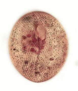
Ventral infraciliature
Description:
Ventral infraciliature of the hymenostome ciliate, Frontonia angusta (Kahl, 1931). Very similar in overall apppearance to F. acuminata (Ehrenberg,1833)Buetschli,1889. F. angusta lacks the anterior apical collection of pigmented granules seen in F. acuminata and its contractile vacuole has 3-4 excretory pores (not visible here).The prominent preoral and postoral sutures are visible. The 3 curved adoral membranelles are seen on the viewer's right of the oral apparatus. The vestibular ciliary rows are seen to the viewer's left of the the oral apparatus.The postoral ciliary field is seen abutting the posterior margin of the peristome to the viewer's right of the postoral suture.Stained by the silver carbonate technique (see Foissner, W. Europ. J. Protistol., 27:313-330;1991).Collected from a freshwater pond near Boise, Idaho.Brightfield.
Included On The Following Pages:
- Life (creatures)
- Cellular (cellular organisms)
- Eukaryota (eukaryotes)
- SAR (Stramenopiles, Alveolates, Rhizaria)
- Alveolata (alveolates)
- Ciliophora (ciliates)
- Intramacronucleata
- Oligohymenophorea
- Peniculida (Peniculid)
- Frontoniidae
- Frontonia
- Frontonia angusta
This image is not featured in any collections.
Source Information
- license
- cc-by-nc
- author
- William Bourland
- provider
- micro*scope
- original
- original media file
- visit source
- partner site
- micro*scope
- ID