cortex
Description:
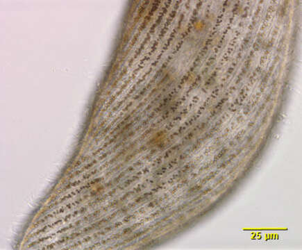
Cortical detail of the marine heterotrich ciliate, Condylostoma arenarium (Spiegel, 1926). The dorsoventrally flattened elongate cell body is very contractile. Contraction is probably mediated by calcium dependent subcortical myonemes and cell extension by interaction of cortical postciliary microtubular ribbons. The broad anterior V-shaped peristome is bordered on the right by a large undulating membrane. An adoral zone of membranelles (AZM) winds from right anterior clockwise around the left margin of the peristome (not visible in this image). Strips of yellowish cortical granules or pigmentocysts separate uniform longitudinal somatic kineties. Pigmentocysts are extrusomes containing toxic substances. They play a role in cell defense against predators and may also function in photoreception. Pigmentocysts are found in other heterotrichs (e.g. Blepharisma and Stentor species). Several cirri are located at the right-most end of the AZM. The long moniliform macronucleus extends along the right cell margin (not visible in this image). No contractile vacuole. Brownish food vacuoles throughout the cytoplasm in this individual contain ingested dinoflagellates (Amphidinium). Collected from a seawater aquarium in Boise, Idaho January 2004. DIC optics.
Included On The Following Pages:
- Life (creatures)
- Cellular (cellular organisms)
- Eukaryota (eukaryotes)
- SAR (Stramenopiles, Alveolates, Rhizaria)
- Alveolata (alveolates)
- Ciliophora (ciliates)
- Postciliodesmatophora
- Heterotrichea
- Heterotrichida
- Condylostomatidae
- Predurostyla
- Predurostyla arenaria
This image is not featured in any collections.
Source Information
- license
- cc-by-nc
- author
- Bill Bourland
- provider
- micro*scope
- original
- original media file
- visit source
- partner site
- micro*scope
- ID