Image of Mcvaughia
Description:
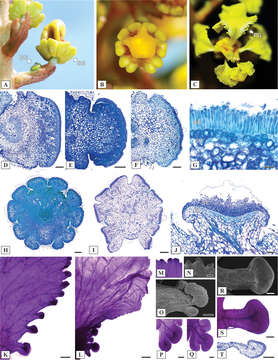
Figure 4. Reproductive morphoanatomy of Mcvaughia species. A inflorescence during development, showing a bracteole gland (BG) and Sepal glands (SG) B ten sepal glands encircling the calyx C Petal glands (PG) along the margin of posterior petal D–F transverse section of bracteole glands in M.sergipana, M.bahiana and M.piauhiensis, respectively G anatomical arrangement of bracteole gland, with a palisade-like secretory epidermis (SE) and secretory parenchyma (SP) H–I transverse section of floral bud and anthesis flower in Mcvaughiabahiana and M.sergipana; calyx gland pair displaced at the anterior sepal J calyx gland structure, showing a secretory epidermis (SE) and vascularized secretory parenchyma (SP) K–L petal glands on the margin of petals in M.sergipana and M.bahiana respectively M–N detail of the petal glands at the apex of the petal limb in M.sergipana, cleared and in SEM image O–Q petal glands positioned at the base, M.bahiana on SEM image, M.bahiana and M.piauhiensis cleared R–T conspicuous and stalked petal glands at the base of M.sergipana, in SEM image, cleared and longitudinal section. Scale bars: 200 μm (D), 150 μm (E–F), 50 μm (G), 500 μm (H–I), 100 μm (J, P–S), 300 μm (L–M), 200 μm (N, T).
Included On The Following Pages:
- Life (creatures)
- Cellular (cellular organisms)
- Eukaryota (eukaryotes)
- Archaeplastida (plants)
- Chloroplastida (green plants)
- Spermatophytes (seed plants)
- Angiosperms (Dicotyledons)
- Eudicots
- Superrosids
- Rosids
- Malpighiales
- Malpighiaceae (Barbados cherry family )
- Mcvaughia
- NO NAME!
This image is not featured in any collections.
Source Information
- license
- cc-by-3.0
- copyright
- Rafael F. Almeida, Isabel R. Guesdon, Marcelo R. Pace, Renata M.S. Meira
- bibliographic citation
- Almeida R, Guesdon I, Pace M, Meira R (2019) Taxonomic revision of Mcvaughia W.R.Anderson (Malpighiaceae): notes on vegetative and reproductive anatomy and the description of a new species PhytoKeys (117): 45–72
- original
- original media file
- visit source
- partner site
- Phytokeys
- ID