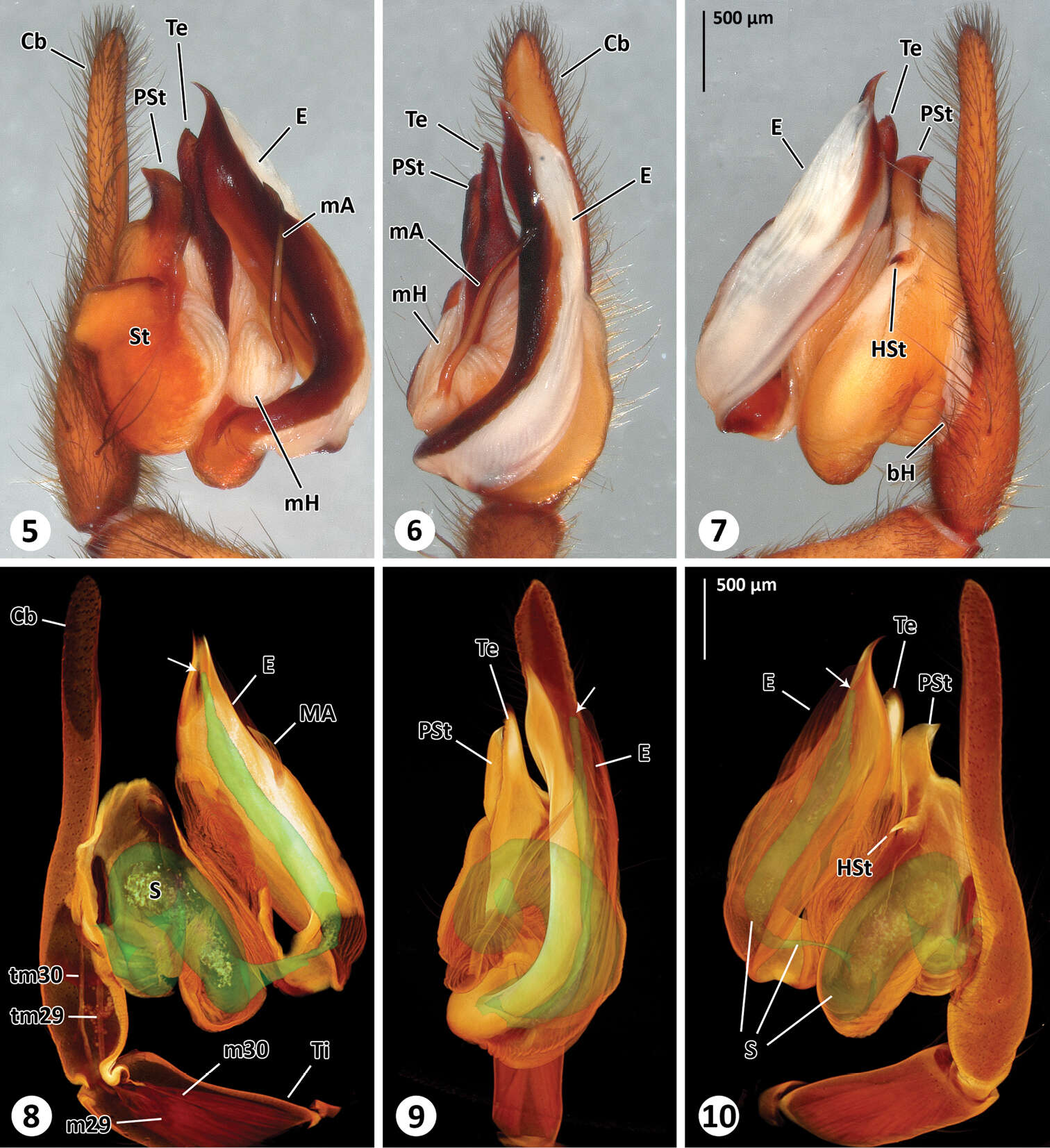
Figure 5–10.Left male palp of Thaida chepu. 5–7 extended focal plane images of male palp in prolateral (5), ventral (6) and retrolateral (7) view 8–10 surface model of the spermophor superimposed on the volume rendering of the male palp to illustrate dimension and shape of the spermophor. The views correspond to Figs 5–7. The cymbium, subtegulum and tegulum are (partly) removed in Fig. 8 to show tendons and muscles. The arrows point to the opening of the embolus.
Video 1.Surface model of the spermophor superimposed on the volume rendering of the male palp. Video available for download in full resolution from http://www.pensoft.net/J_FILES/1/articles/6021/export.php_files/Michalik_Ramirez_Video_1.avi.