-
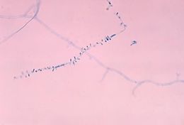
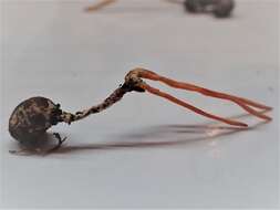
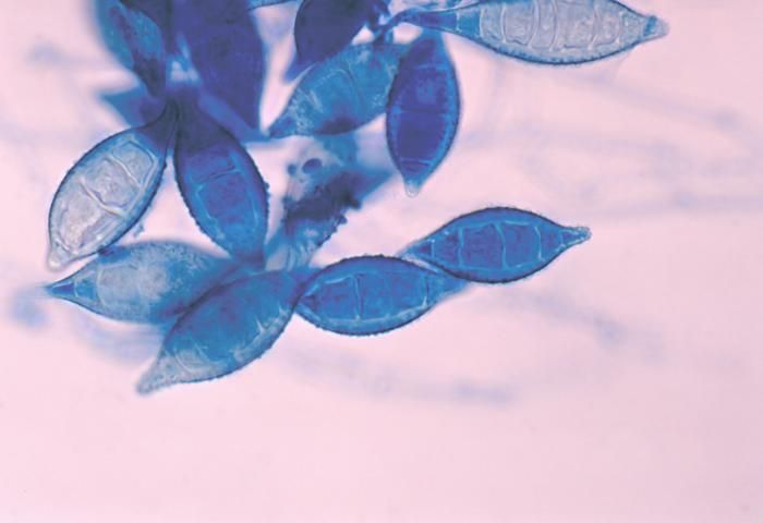
This photomicrograph shows a number of Arthroderma otae, formerly Nannizzia otae, fungal macroconidia.Created: 1978
-
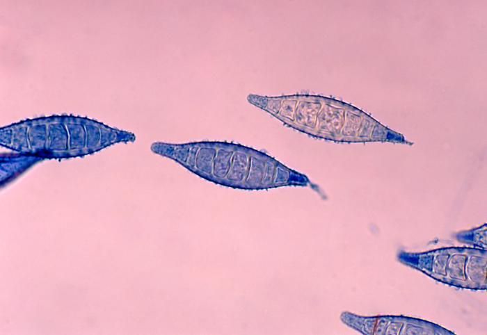
This photomicrograph shows a number of Arthroderma otae, formerly Nannizzia otae, fungal macroconidia.Created: 1978
-
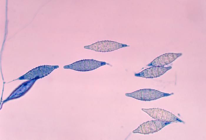
This photomicrograph shows a number of Arthroderma otae, formerly Nannizzia otae, fungal macroconidia.Created: 1978
-
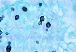
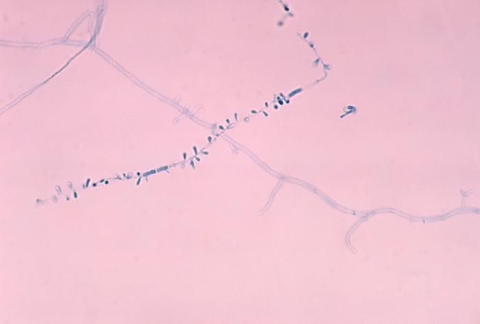
This photomicrograph shows a number of microconidia of the fungus Arthroderma otae, formerly Nannizzia otae.Created: 1978
-
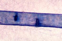
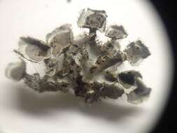
This micrograph reveals the hair perforation caused by the fungus Arthroderma otae, formerly Nannizzia otae.Created: 1978
-
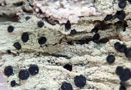
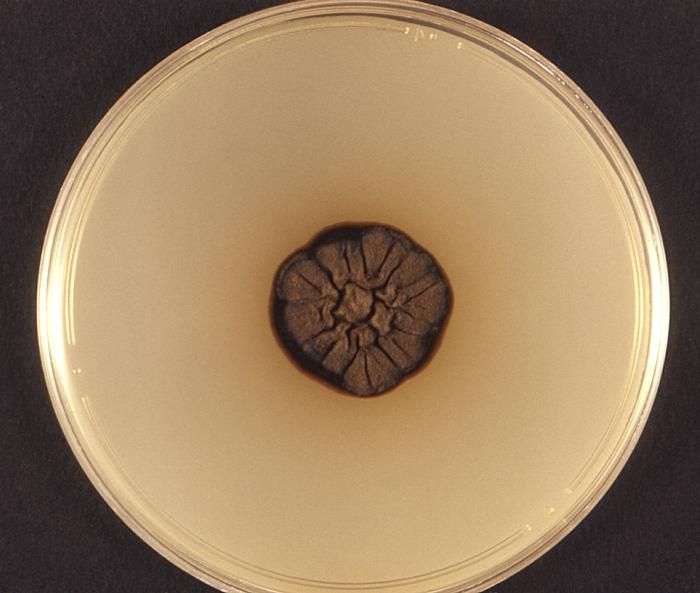
From a frontal view, this photograph depicts a single colony of Ochroconis humicola, fungi, formerly known as Scolecobasidium humicola, which had been isolated from a specimen of infected fish.Created: 1974
-
Note the histopathologic changes seen in histoplasmosis due to Histoplasma capsulatum var. duboisii.Created: 1972
-
Note the histopathologic changes seen in histoplasmosis due to Histoplasma capsulatum var. duboisii.Created: 1972
-
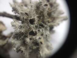
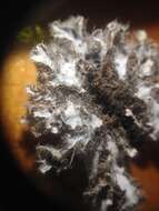
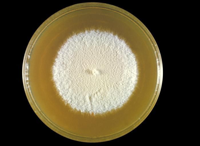
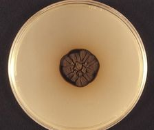
This was a plate culture of the fungus Chrysosporium keratinophilum.Created: 1963
-
-
-
-
-
-
-
-
-
-
-
-
-
-
-