-
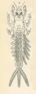
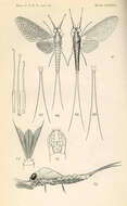
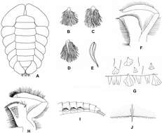
Pentagenia vittigera Walsh; nymph. Nymph from above, with the gills on the right pushed aside to show the markings of the abdomen
-
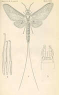
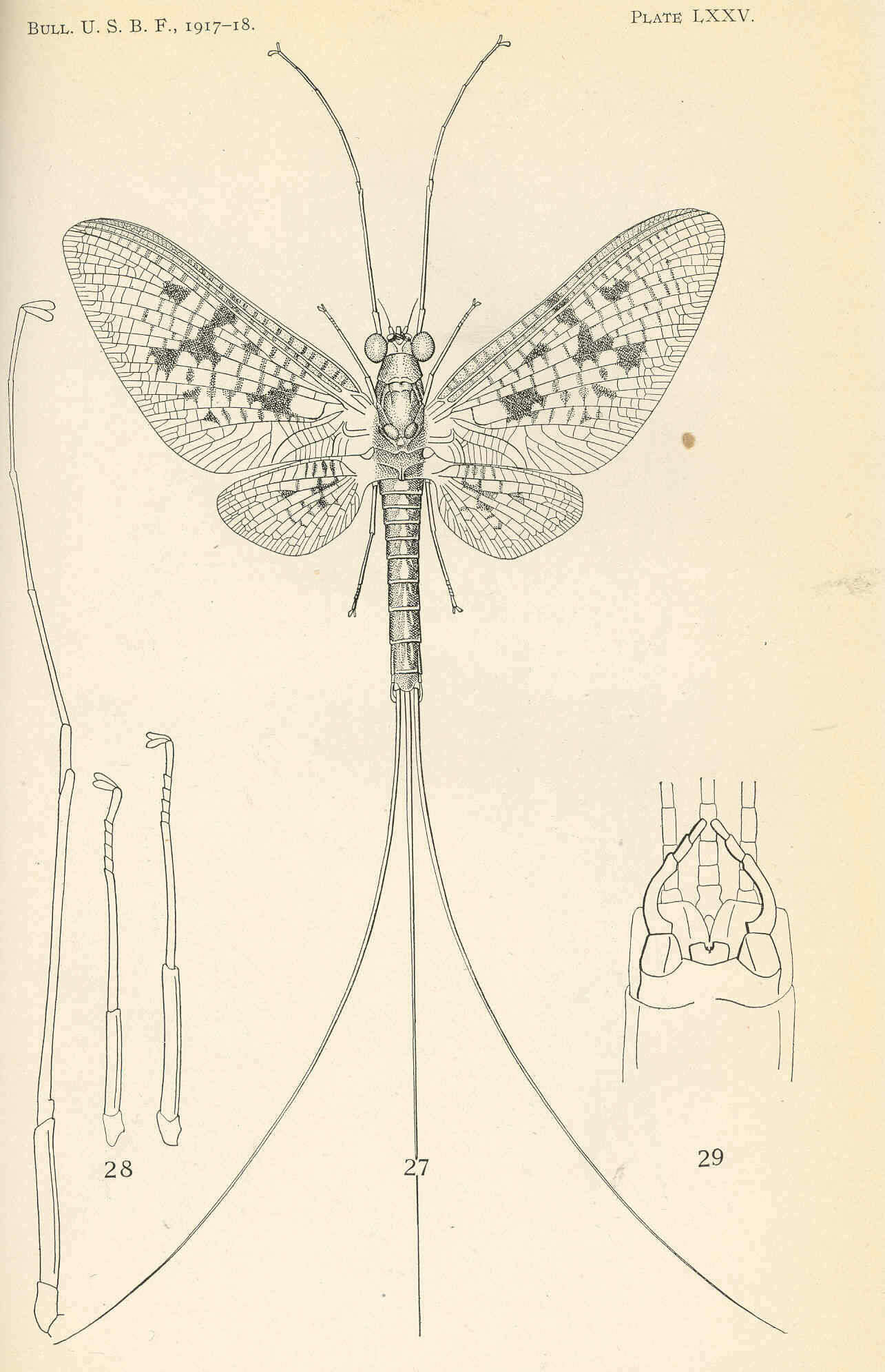
Ephemera varia Eaton; adult. Adult male 27; Legs of one side of the same 28; End of abdomen of the same from beneath 29
-
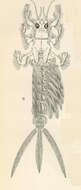
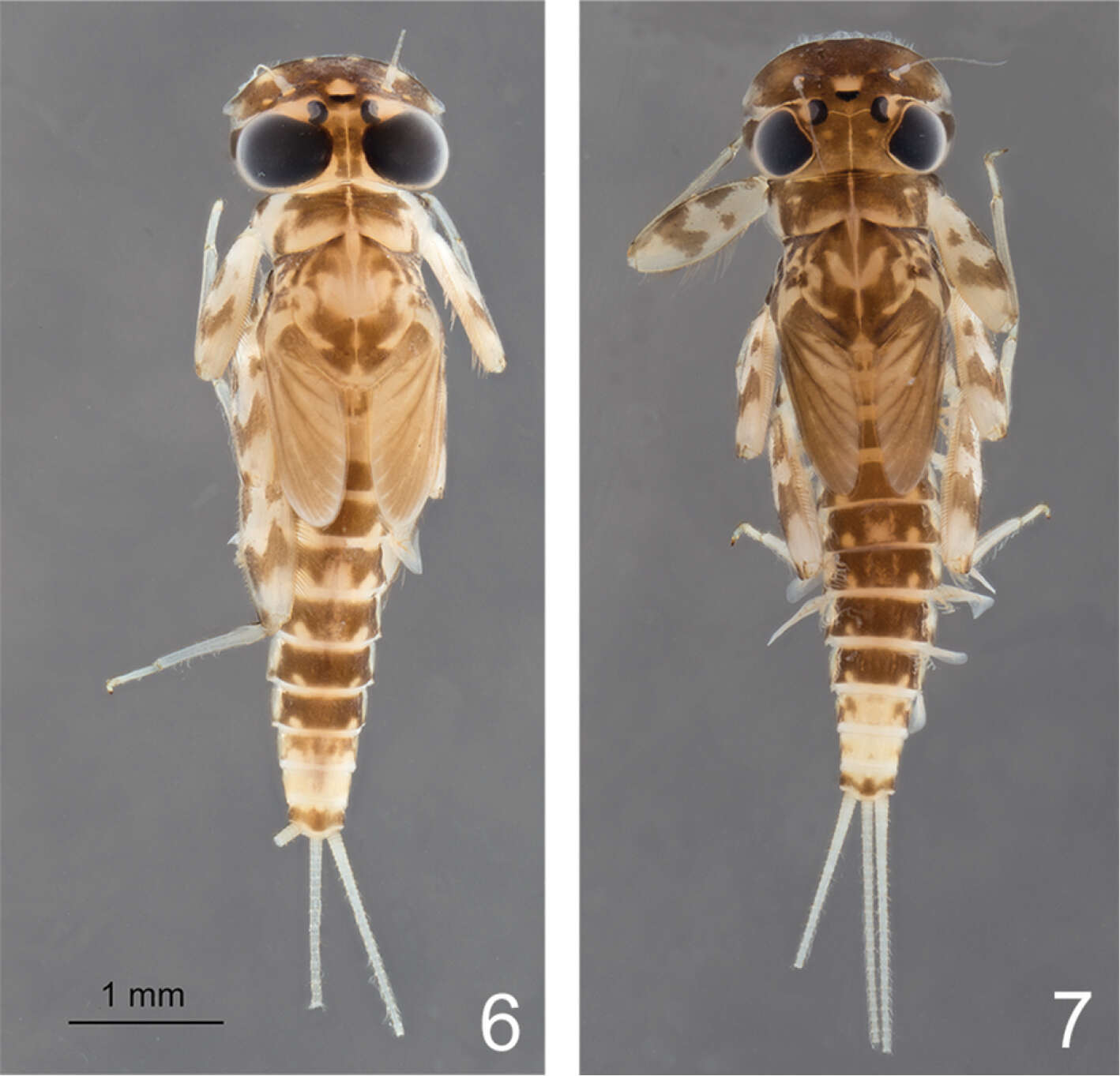
Hexagenia nymphs. Nymph of H. bilineata Say, dorsal view. The hind feet are turned forward for comparison with the other feet, and the gills on the right are moved aside to show the markings on the abdomen. The 3-branched, rudimentary gills on the first abdominal segment are abnormal
-
Boonsatien Boonsoong, Dietrich Braasch
Zookeys
Figure 1.A Habitus of Epeorus aculeatus Braasch, 1990 B lamellae of gills 7 of Epeorus khayengensis Boonsoong & Braasch, 2010 C–E ventral view of abdomen (C), abdominal gills 1 (D) and abdominal terga of (E) Epeorus thailandensis sp. n. F abdominal terga of Epeorus unicornutus Braasch, 2006 G abdominal terga of Epeorus khayengensis Boonsoong & Braasch, 2010 H–J abdominal terga (H), lamellae of gills 1 (I) and tergum VII (J) of Epeorus inthanonensis Braasch & Boonsoong, 2010.
-
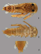
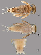
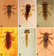
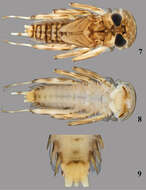
Figures 1–3.Thalerosphyrus determinatus (Walker, 1853). 1 Habitus in dorsal view 2 Habitus in ventral view 3 Detail of abdominal segments VI–IX in ventral view.
-
Figures 1–4.Rhithrogeniella ornata Ulmer, 1939. 1 Genitalia of the male imago (holotype) in ventral view 2 Foreleg of a male subimago (paratype) 3 Hindleg of a male subimago (paratype) 4 Penis lobes of a male subimago (paratype): plain line, cuticular structures of the subimago; dotted line, outline of the imago penis lobes.
-
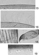
Pentagonia vittigera Walsh; adult. Adult mail of the quadripunctata form 15; Legs of one side 16; End of abdomen of male from beneath 17; End of abdomen of female from beneath 18
-
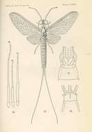
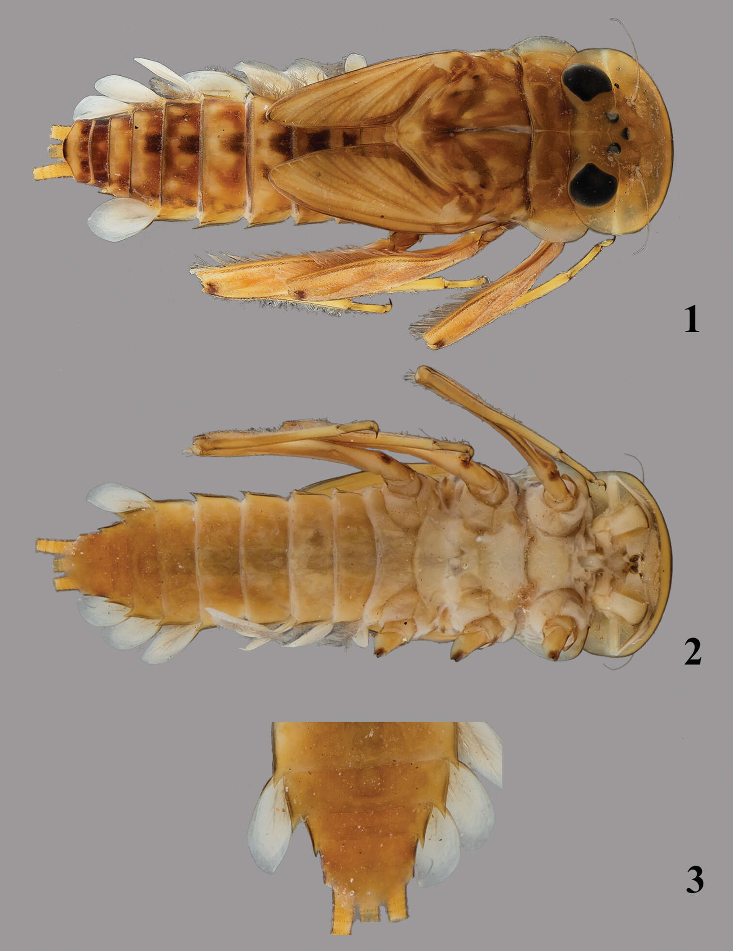
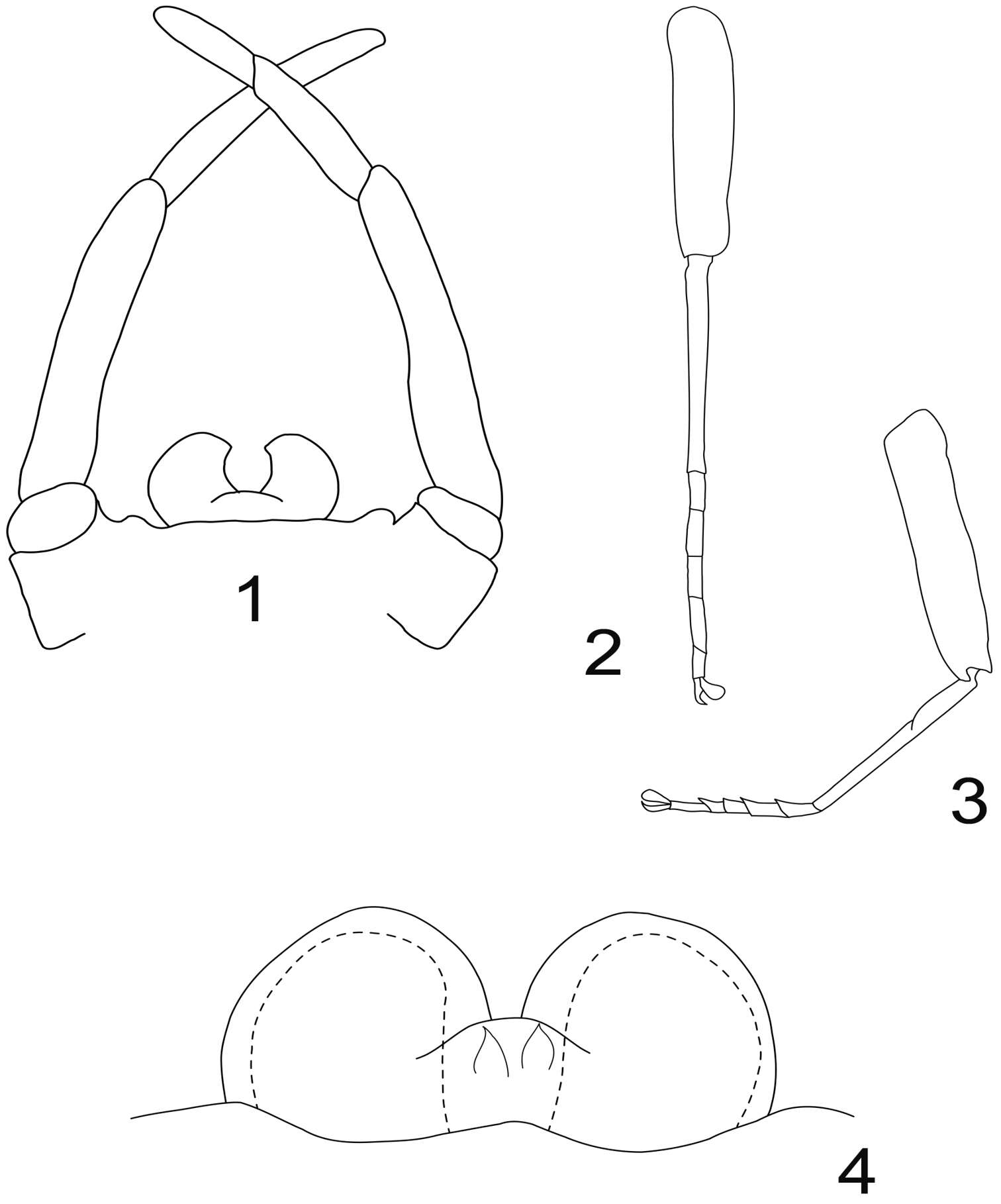
Hexagenia bilineata Say; adult. Adult male (segmentation of the tails omitted...) 1; Legs of one side, fore, middle, and hind from left to right... 2; end of the abdomen of the male from beneath, showing forceps, penes, and base of tails; middle tail rudimentary
-
Boonsatien Boonsoong, Dietrich Braasch
Zookeys
Figure 5.A–B General outline (A) and micropyle (B) of the egg of Epeorus khayengensis Boonsoong & Braasch, 2010 C-D General outline (C) and micropyle (D) of the egg of Rhithrogena siamensis Braasch & Boonsoong, 2009. Scale bars 20 µm for A and C; 5 µm for B and D.
-
Figures 4–6.Thalerosphyrus sinuosus (Navás, 1933). 4 Habitus in dorsal view 5 Habitus in ventral view 6 Detail of abdominal segments VI–IX in ventral view.
-
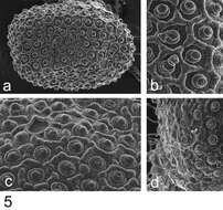
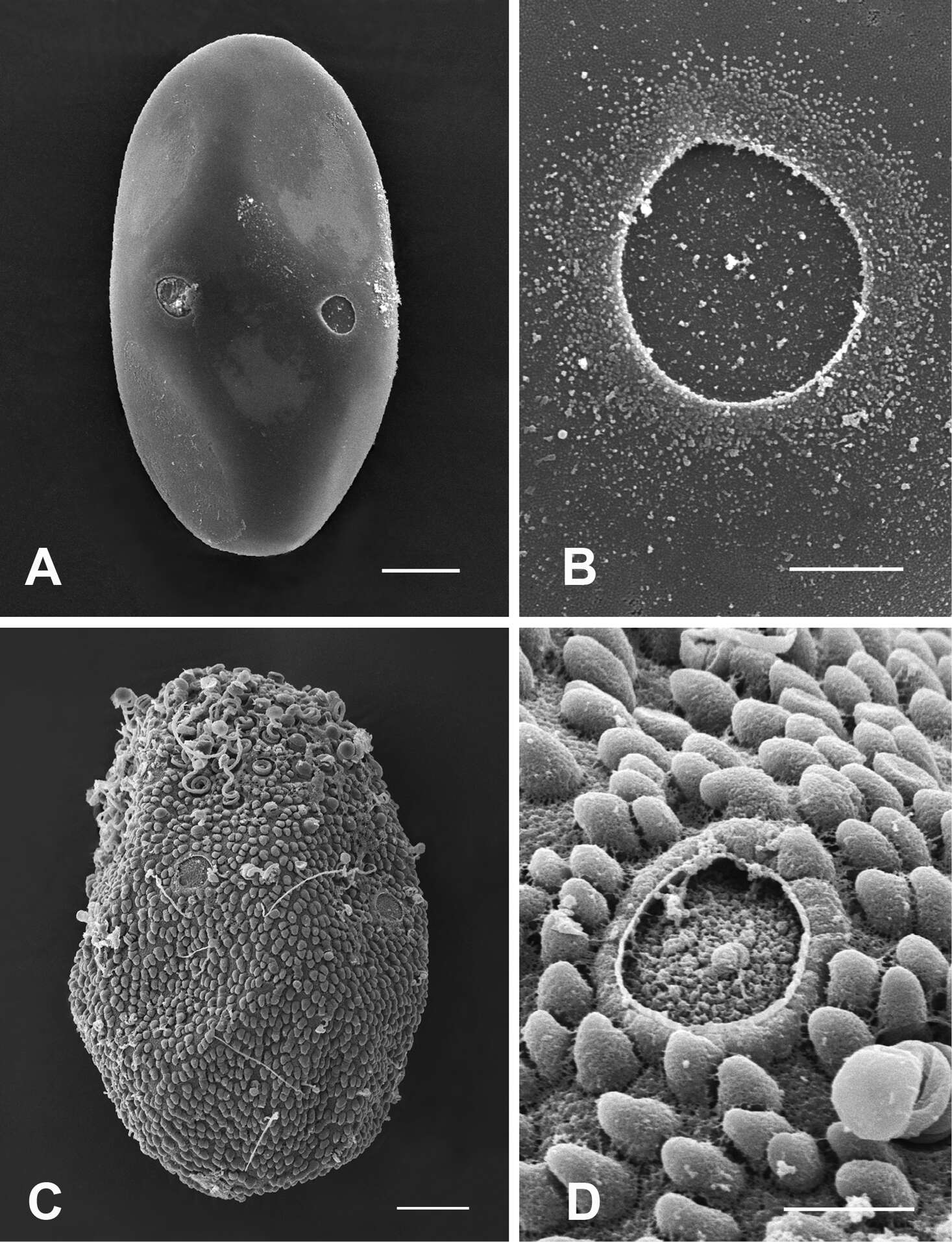
Figure 5.Rhithrogeniella ornata Ulmer, 1939, SEM pictures of egg structures. 5a Egg extracted from a female subimago paratype from Padang, Sumatra 5b Details of the chorionic structure of a female nymph from Ombilin River, Sumatra 5c Details of the chorionic structure and micropyle of a female subimago paratype from Buitenzorg [Bogor], Java 5d chorionic surface of the female allotype from Buitenzorg [Bogor], Java.
-
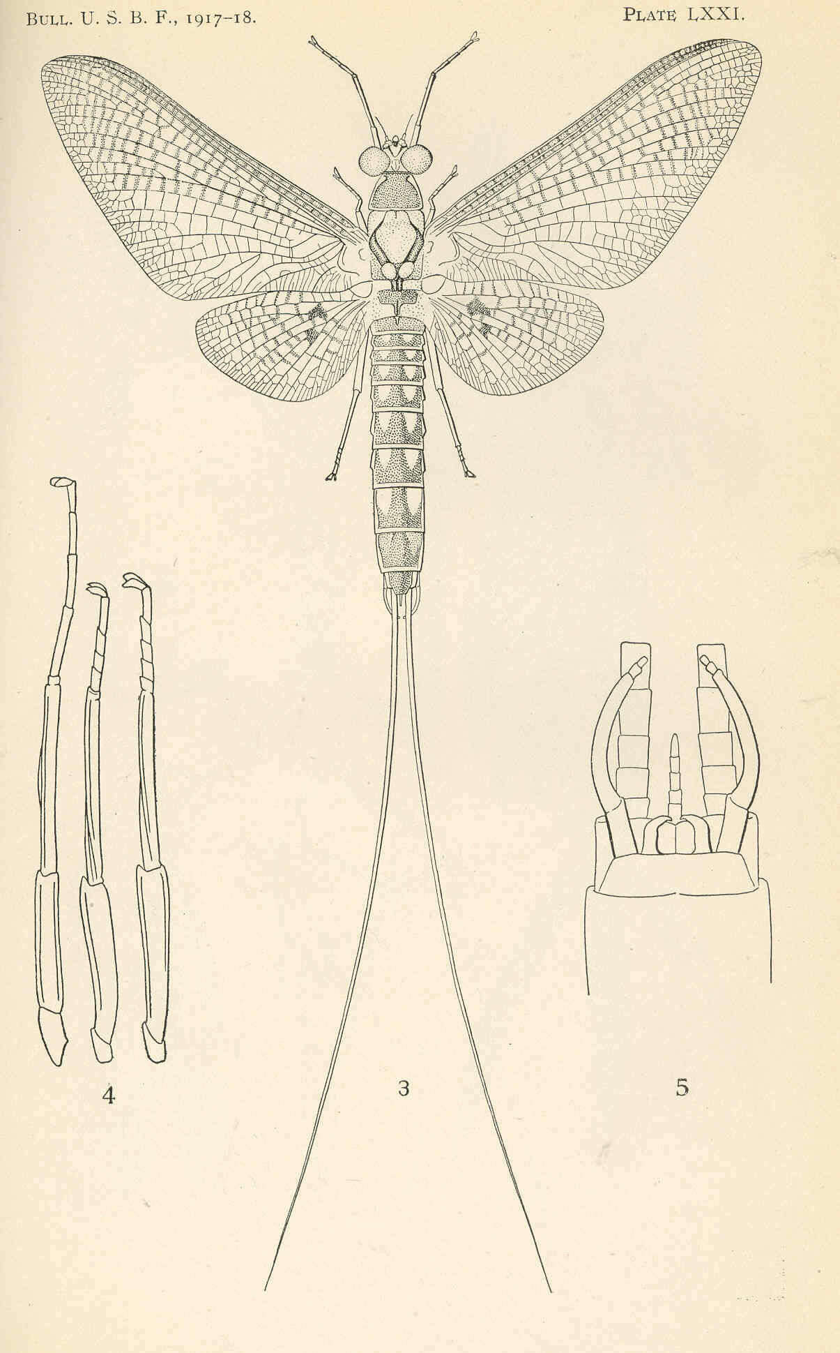
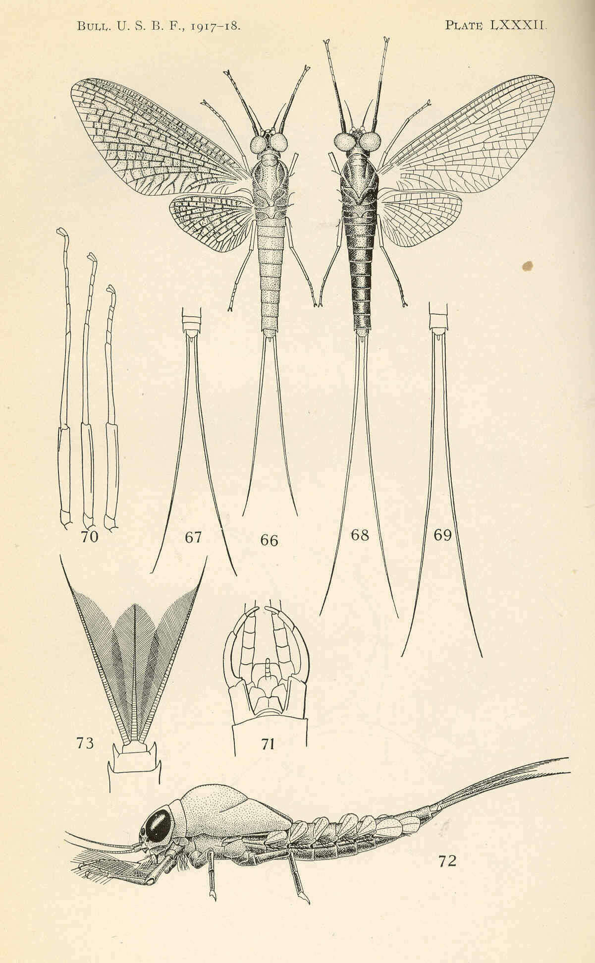
Chirotenetes siccus Walsh. Female subimago 66; Sketch of tails of male subimago to show their relative length 67; Female imago 68; Sketch of tails of male imago to show their relative length 69; legs of one side of male imago 70; End of abdomen of adult male from beneath 71; Nymph from the side. Note the plancton-gathering fringes of the fore legs 72; Tail fin of the nymph; outer tails fringed only on the inner side 73
-
Boonsatien Boonsoong, Dietrich Braasch
Zookeys
Figure 9.A Habitus of Asionurus namnaoensis Braasch & Boonsoong, 2010 B habitus of Asionurus primus Braasch & Soldán, 1986 Chabitus of Compsoneuria thienemanniUlmer, 1939 Dhabitus of Epeorus khayengensis Boonsoong & Braasch, 2010 E habitus of Rhithrogena tonkinensis Soldán & Braasch, 1986 F habitus of Thalerosphyrus sinuosus Navás, 1933.
-
Figures 7–9.Thalerosphyrus lamuriensis Sartori, 2014. 7 Habitus in dorsal view 8 Habitus in ventral view 9 Detail of abdominal segments VI–IX in ventral view.
-
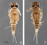
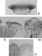
Figures 6–7.Rhithrogeniella ornata Ulmer, 1939. 6 Male nymph 7 Female nymph with slight color variations.
-
Boonsatien Boonsoong, Dietrich Braasch
Zookeys
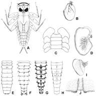
Figure 2.A Ventral view of abdomen of Rhithrogena siamensis Braasch & Boonsoong, 2009 B–E lamellae of gills 1 (B), 3 (C), 5 (D) and 7 (E) of Trichogenia maxillaris Braasch & Soldán, 1988 F ventral view of left maxilla of Trichogenia maxillaris Braasch & Soldán, 1988 G bristles on dorsal face of abdominal terga of Trichogenia maxillaris Braasch & Soldán, 1988 H ventral view of left maxilla of Compsoneuria langensis Braasch & Boonsoong, 2010 I-J abdominal terga (I) and tergum VII (J) of Notacanthurus baei Braasch & Boonsoong, 2009.
-
Figures 16–22.Mouthparts structure of Thalerosphyrus determinatus (20), Thalerosphyrus sinuosus (16, 21) and Thalerosphyrus lamuriensis (17, 18, 19, 22). 16–17 Hemi-labrum 18 Left mandible 19 Right mandible 20–22 Labial glossa.
-
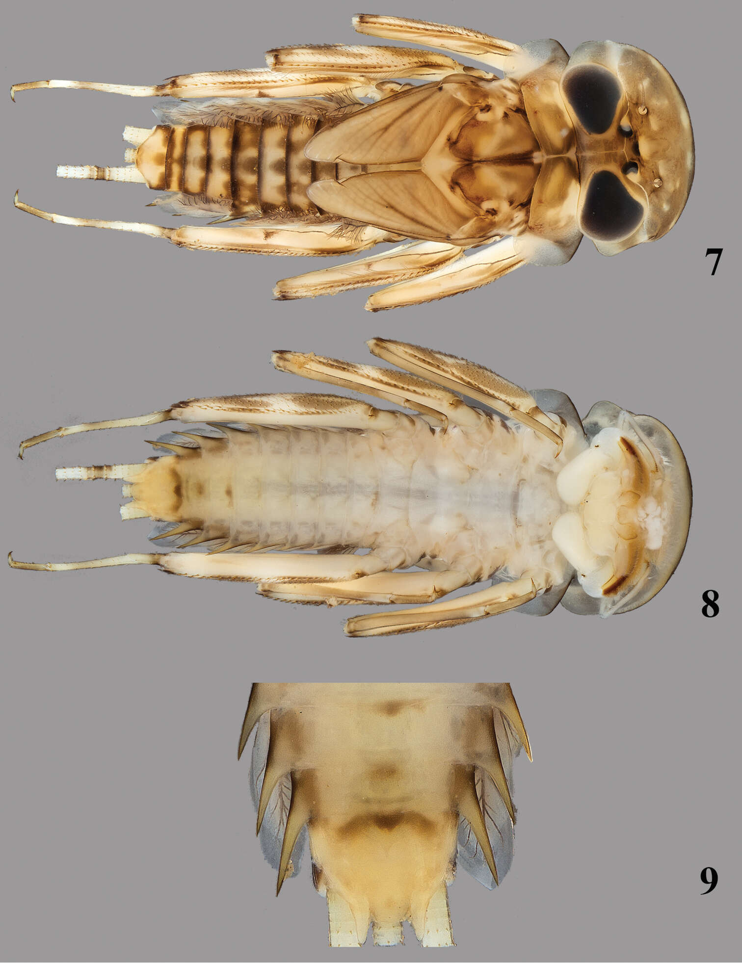
Figures 8–11.Rhithrogeniella ornata Ulmer, 1939, nymphal mouthparts. 8 Labrum in dorsal view 9 Left glossae and paraglossae of the labium 10 Detail of the glossae from 9 11 Hypopharynx, ventral view lingua and left superlingua.
-
Boonsatien Boonsoong, Dietrich Braasch
Zookeys
Figure 5.A–B General outline (A) and micropyle (B) of the egg of Epeorus khayengensis Boonsoong & Braasch, 2010 C-D General outline (C) and micropyle (D) of the egg of Rhithrogena siamensis Braasch & Boonsoong, 2009. Scale bars 20 µm for A and C; 5 µm for B and D.
-
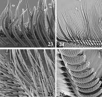
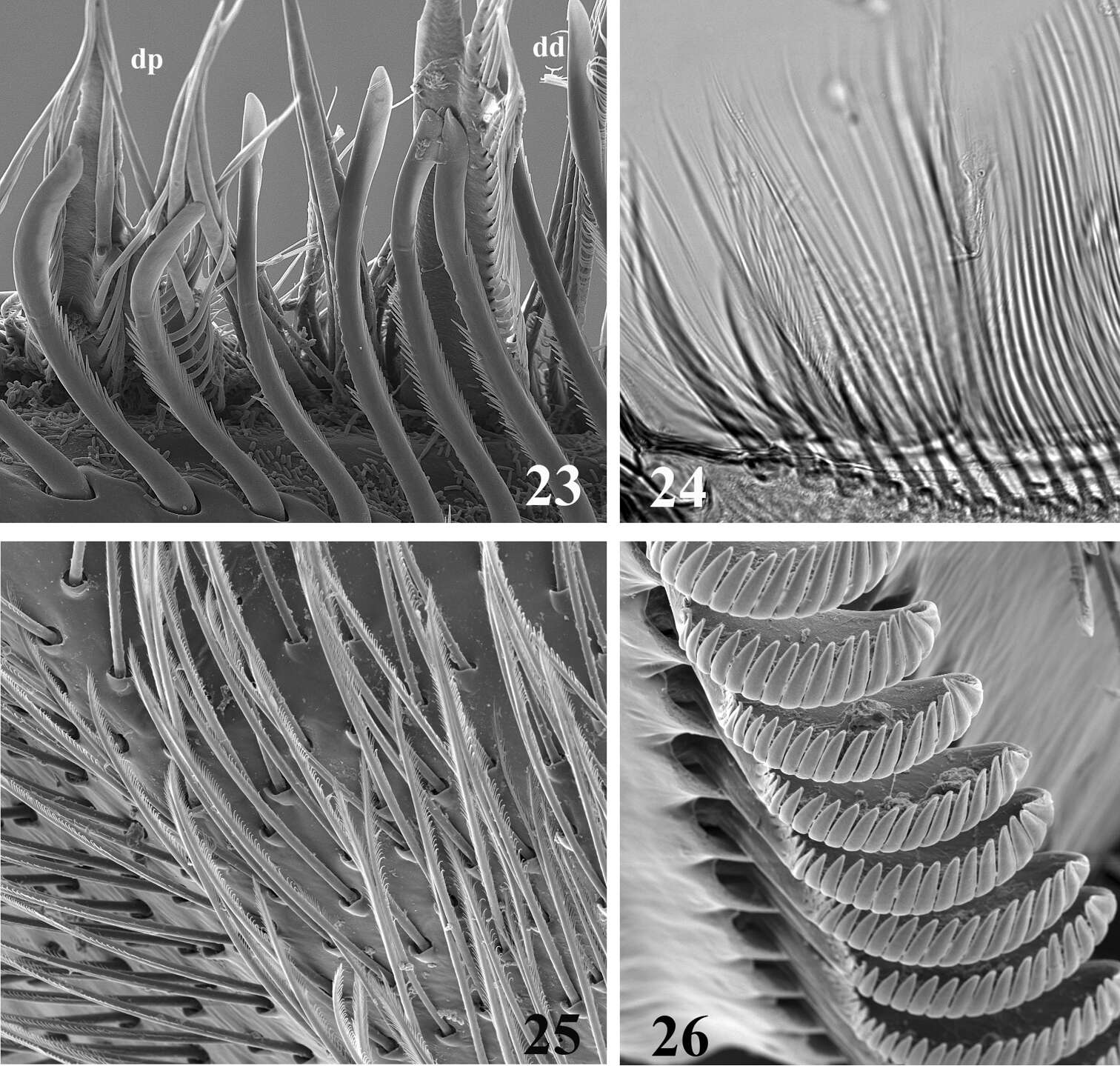
Figures 23–26.SEM (23, 25, 26) and optic (24) pictures of maxillar structure. 23–24 Dentisetae of Thalerosphyrus lamuriensis dp: proximal dentisetae, dd distal dentisetae 25 Scattered setae on the ventral face of the galea-lacinia of Thalerosphyrus sinuosus 26 Comb-shape setae on the crown of the galea-lacinia of Thalerosphyrus sinuosus.
-
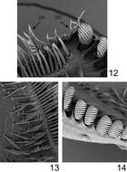
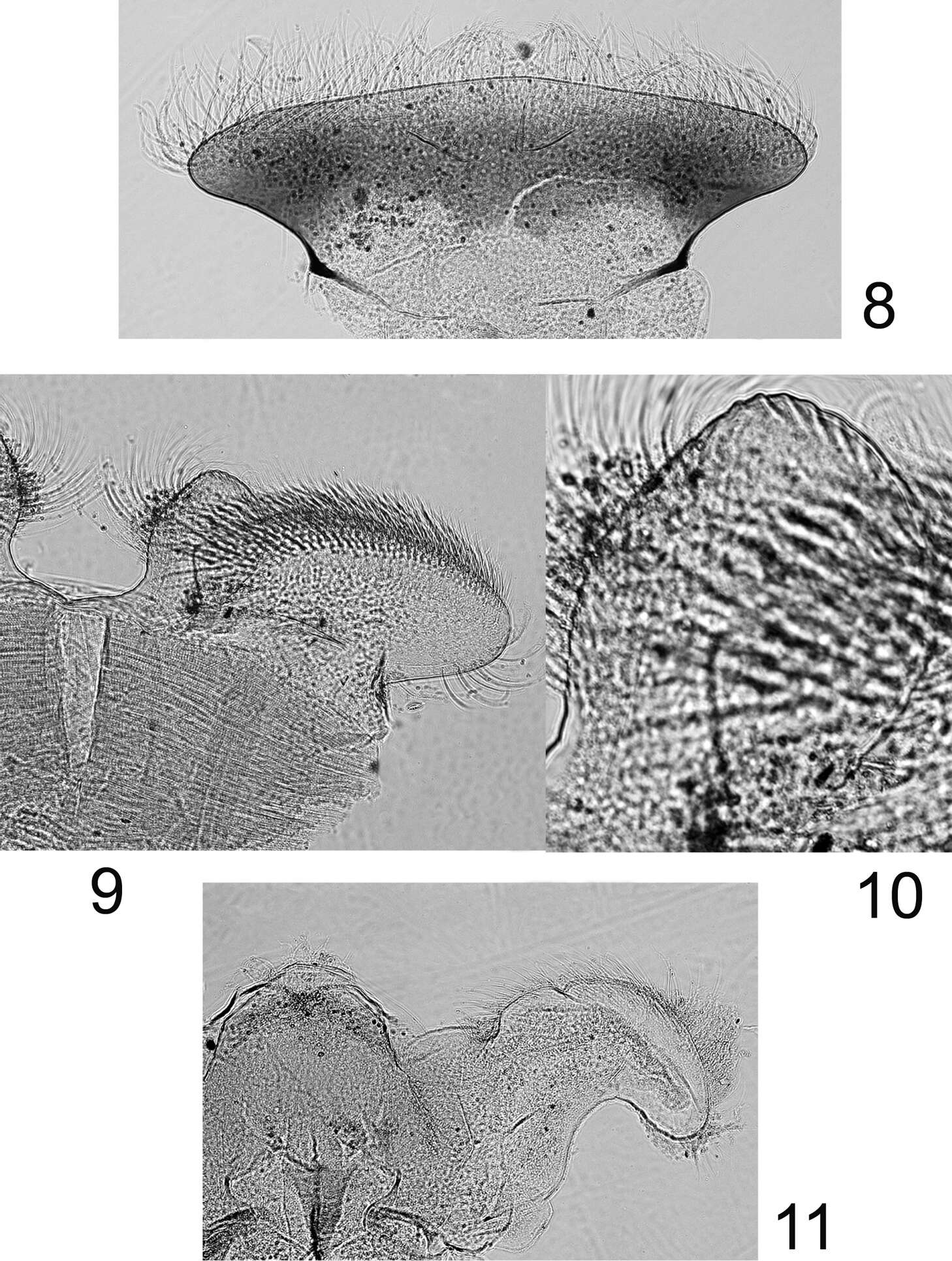
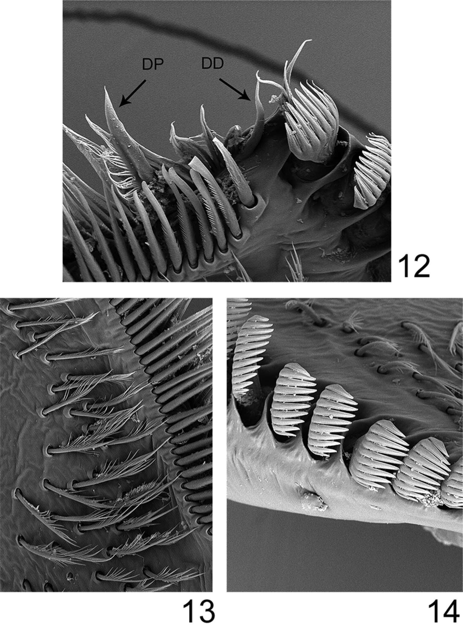
Figures 12–14.Rhithrogeniella ornata Ulmer, 1939, SEM pictures of the maxilla. 12 Dentisetae (DP: proximal dentiseta, DD: distal dentiseta) 13 Fimbriate setae on the ventral surface 14 Comb-shape setae on the crown of the galea-lacinia.
-
Boonsatien Boonsoong, Dietrich Braasch
Zookeys
Figure 2.A Ventral view of abdomen of Rhithrogena siamensis Braasch & Boonsoong, 2009 B–E lamellae of gills 1 (B), 3 (C), 5 (D) and 7 (E) of Trichogenia maxillaris Braasch & Soldán, 1988 F ventral view of left maxilla of Trichogenia maxillaris Braasch & Soldán, 1988 G bristles on dorsal face of abdominal terga of Trichogenia maxillaris Braasch & Soldán, 1988 H ventral view of left maxilla of Compsoneuria langensis Braasch & Boonsoong, 2010 I-J abdominal terga (I) and tergum VII (J) of Notacanthurus baei Braasch & Boonsoong, 2009.
-
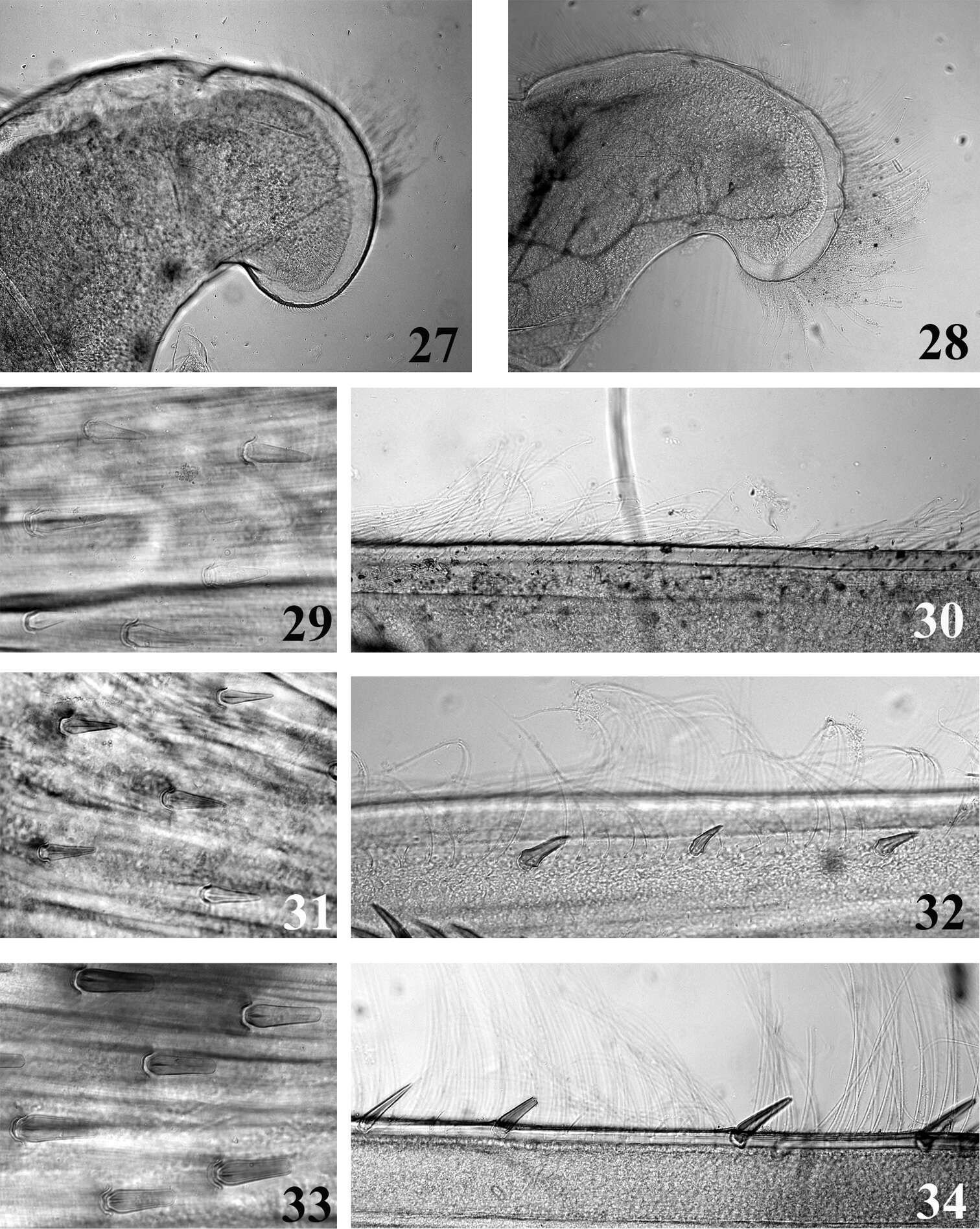
Figures 27–34.Mouthpart (27–28) and thoracic (29–34) structures of Thalerosphyrus determinatus (27, 29, 30), Thalerosphyrus sinuosus (31, 32) and Thalerosphyrus lamuriensis (28, 33, 34). 27–28 Apex of superlingua of hypopharynx 29, 31, 33 Bristles on the dorsal face of hind femur 30, 32, 34 Outer margin of hind tibia.
-
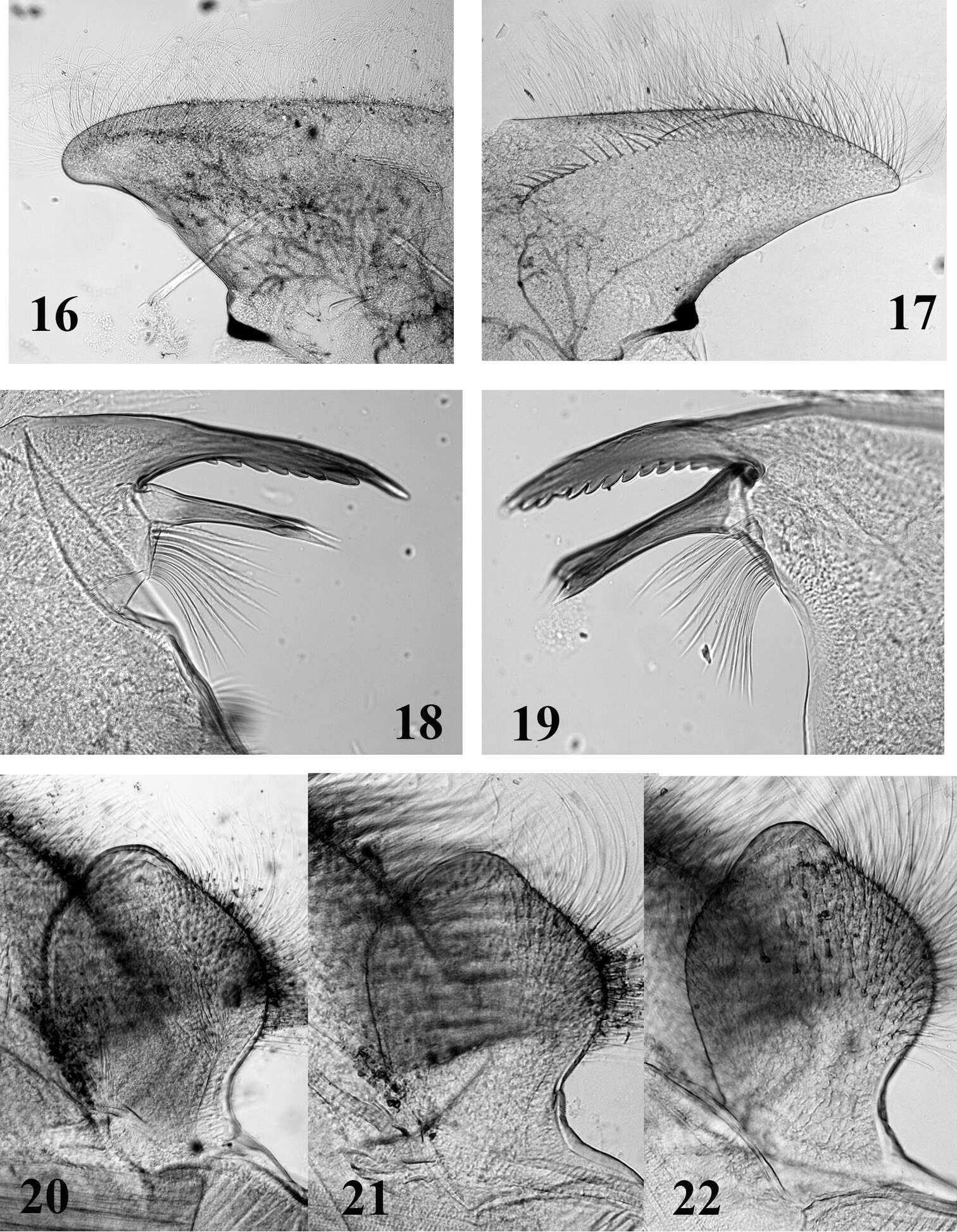
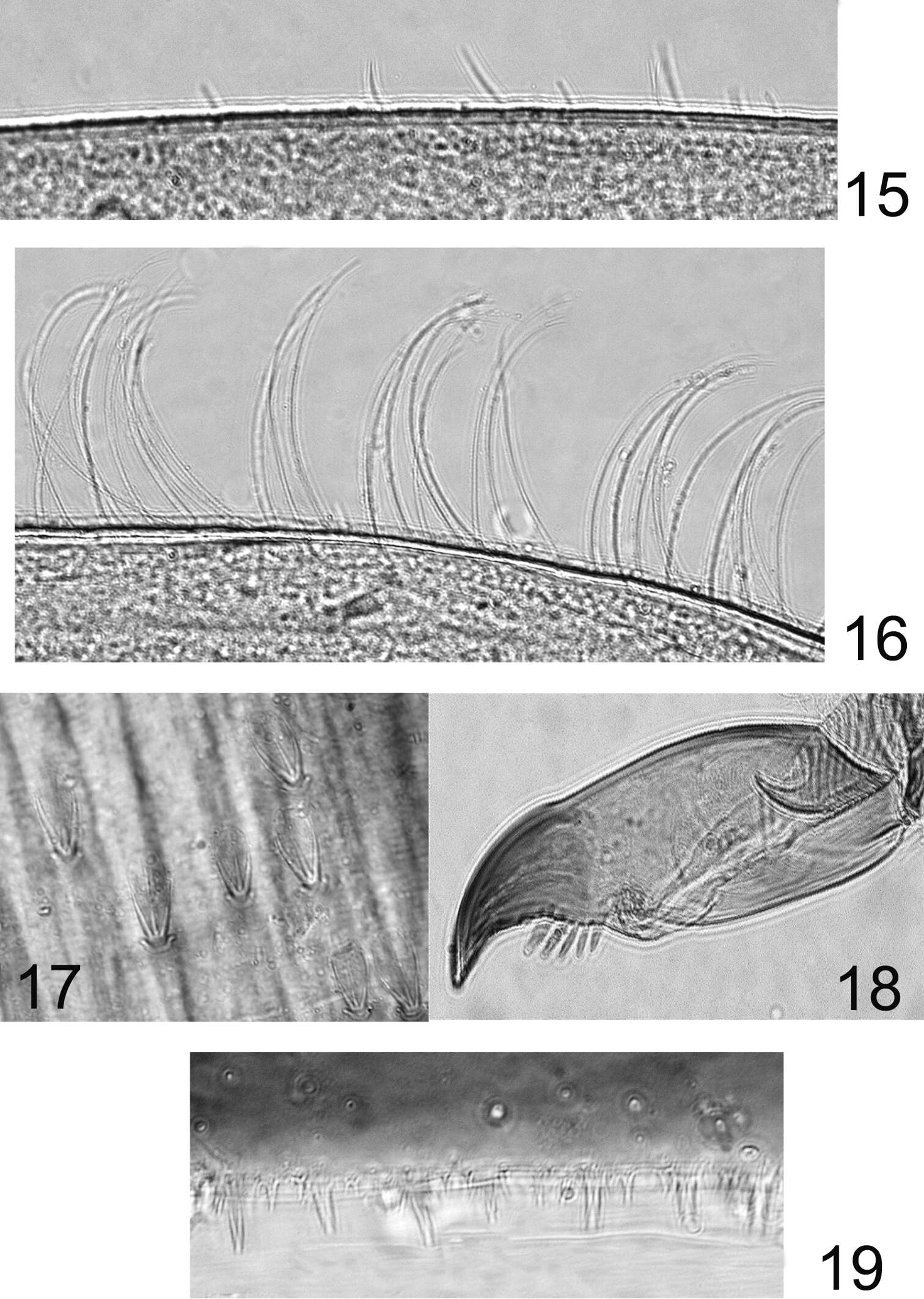
Figures 15–19.Rhithrogeniella ornata Ulmer, 1939. 15 Outer margin of the fore tibia 16 Outer margin of the hind tibia 17 Bristles on the dorsal surface of hind femur 18 Tarsal claw 19 Posterior margin of tergite V.