-
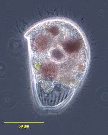
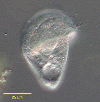
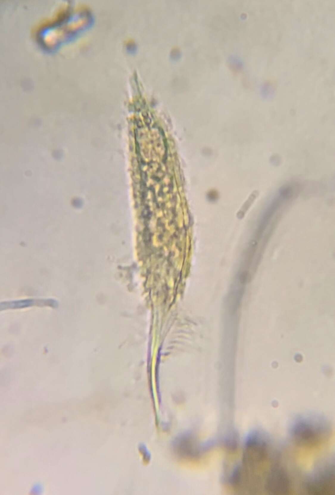
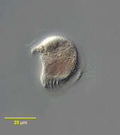
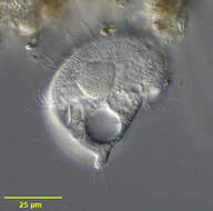
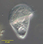
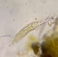
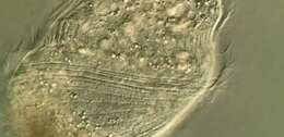
Portrait (anterior oblique view) of the metopid ciliate, Brachonella galeata (Kahl 1927 [Metopus galeatus]; Jankowski, 1964). The anterior half of the cell is broadly helmet-shaped. The posterior is bluntly conical. The peristome encircles the body at the junction of the anterior and posterior halves of the cell, the origin lying just anterior to the termination on the same longitudinal line. The peristome terminates in a posterior cytostome. There is an adoral zone of membranelles on the left margin of the peristome. The posterior part of the AZM is seen in this image. The somatic cilia are long and sparse. There is a tuft of longer cilia at the posterior end. A single ellipsoid macronucleus is located centrally or anteriorly (not seen well here). There is a single irregularly shaped contractile vacuole at the posterior end. There are purple sulfur bacteria visible in food vacuoles. B. galeata is anaerobic. Collected from anoxic sediment of slow-moving freshwater stream near Boise, Idaho in April 2004. DIC optics.
-
Metopus fuscus (Kahl,1927). Stained by the silver carbonate technique (see Foissner, W. Europ. J. Protistol., 27:313-330;1991).Brightfield.
-
Dorsal view of the infraciliature of Metopus hasei (SONDHEIM,1929).The tuft of long caudal cilia is seen here.From a rewetted soil sample collected from the margin of a eutrophic freshwater pond near Boise, Idaho. Stained by the silver carbonate technique (see Foissner, W.Europ. J. Protistol.27:313-330;1991).Brightfield.
-
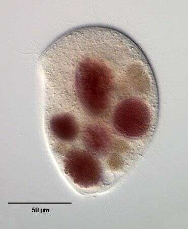
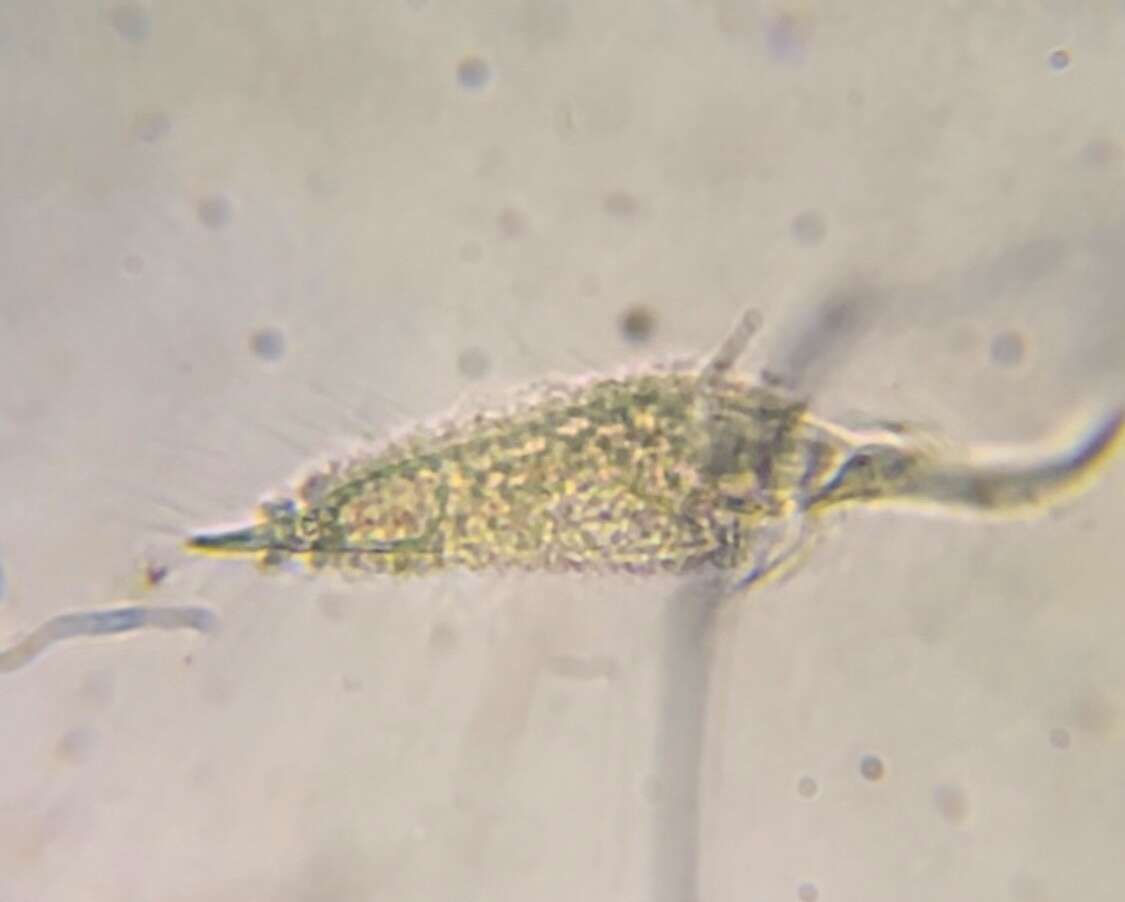
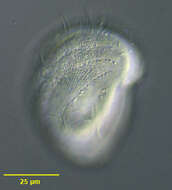
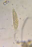
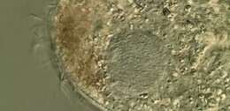
Portrait (coronal section) of the metopid ciliate, Brachonella galeata (Kahl 1927 [Metopus galeatus]; Jankowski, 1964). The anterior half of the cell is broadly helmet-shaped. The posterior is bluntly conical. The peristome encircles the body at the junction of the anterior and posterior halves of the cell, the origin lying just anterior to the termination on the same longitudinal line. The peristome terminates in a posterior cytostome. There is an adoral zone of membranelles on the left margin of the peristome. The somatic cilia are long and sparse. There is a tuft of longer cilia at the posterior end. A single ellipsoid macronucleus is located centrally or anteriorly . There is a single irregularly shaped contractile vacuole at the posterior end. There are purple sulfur bacteria visible in food vacuoles. B. galeata is anaerobic. Collected from anoxic sediment of slow-moving freshwater stream near Boise, Idaho in April 2004. DIC optics.
-
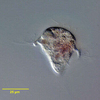
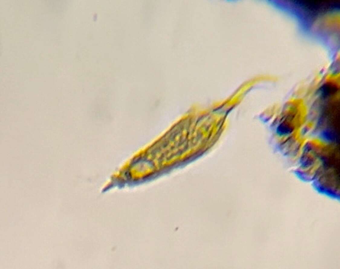
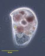
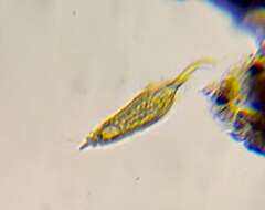
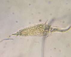
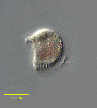
Portrait of Metopus spinosus a polysaprobic ciliate (ventral view). Synonymous with Metopus caudatus. The body is elongate and slightly flattened dorsoventrally. The rounded anterior is twisted to the left and the posterior is drawn out to a tapered point. There is an anterior apical aggregate of refractile granules typical of metopids. Somatic kineties are longitudinal and uniform. The peristome slants obliquely across the ventral surface. There is a prominent adoral zone of membranelles on its left and several kineties of longer cilia along its right margin. The cytostome is at the right posterior end of the peristome. The large ellipsoid central macronucleus is not seen in this image. There is a posterior contractile vacuole.
-
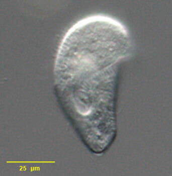
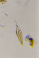
Metopus violaceus (KAHL, 1927). Phase contrast.
-
Metopus violaceus (KAHL, 1927). DIC.
-
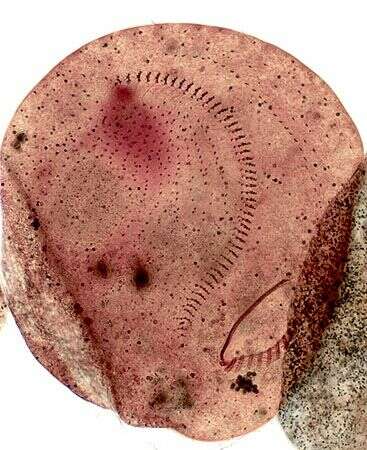
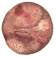
Infraciliature of Metopus violaceus (KAHL, 1927). Stained by the silver carbonate technique (see Foissner, W. Europ. J. Protistol., 27:313-330;1991).Brightfield
-
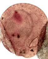
Infraciliature of Metopus violaceus (KAHL, 1927). Stained by the silver carbonate technique (see Foissner, W. Europ. J. Protistol., 27:313-330;1991). Brightfield.
-
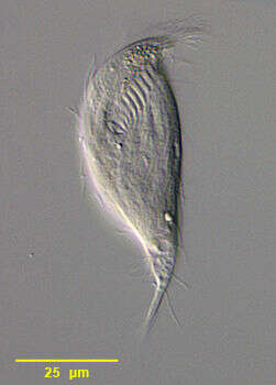
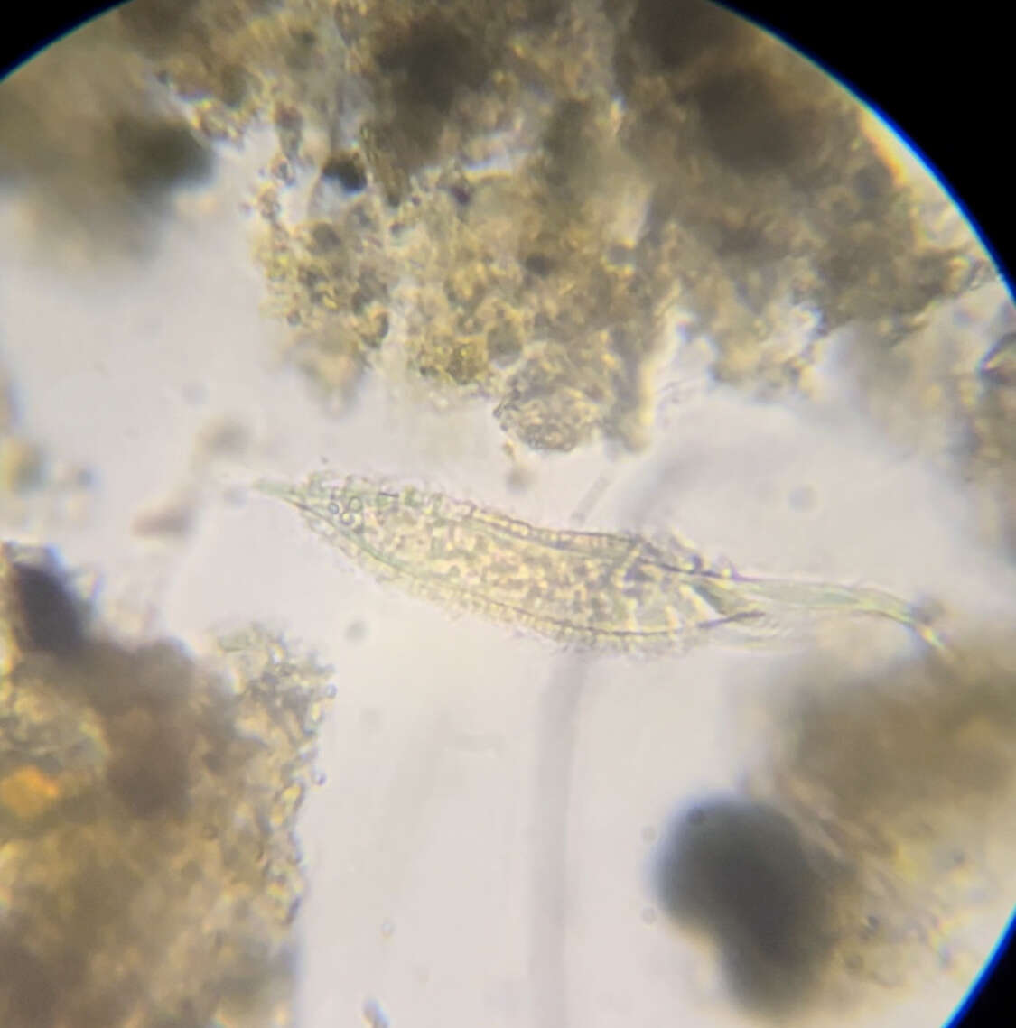
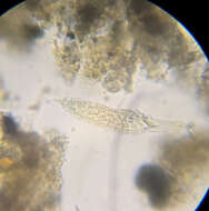
Ventral view of Metopus striatus (synonyms include M. bacillatus, M. gibbus, M. pullus and M. acuminatus among others), a colorless metopid ciliate. The body is broadly domed anteriorly. The posterior end is bluntly tapered or may terminate in a short finger-like process. The oblique peristome with its adoral zone of membranelles terminates at the oral aperture in the posterior 1/3 of the body (seen well here). The large spherical macronucleus is located in the midbody. There is one adjacent micronucleus. The sparse somatic kineties are longitudinal coursing to the left around the anterior dome. There is a longer tuft of caudal cilia. The anterior refractile granules typical of metopids are much less conspicuous in this species. There is a prominent layer of subcortical extrusomes. A single contractile vacuole is located posteriorly. Collected from polysaprobic freshwater pond near Boise, Idaho September 2003. DIC optics.
-
Optical section through Metopus striatus (synonyms include M. bacillatus, M. gibbus, M. pullus and M. acuminatus among others), a colorless metopid ciliate. The body is broadly domed anteriorly. The posterior end is bluntly tapered or may terminate in a short finger-like process. The oblique peristome with its adoral zone of membranelles terminates at the oral aperture in the posterior 1/3 of the body. The large spherical macronucleus is located in the midbody. There is one adjacent micronucleus. The sparse somatic kineties are longitudinal coursing to the left around the anterior dome. There is a longer tuft of caudal cilia. The anterior refractile granules typical of metopids are much less conspicuous in this species. There is a prominent layer of subcortical extrusomes. A single contractile vacuole is located posteriorly. Collected from polysaprobic freshwater pond near Boise, Idaho September 2003. DIC optics.
-
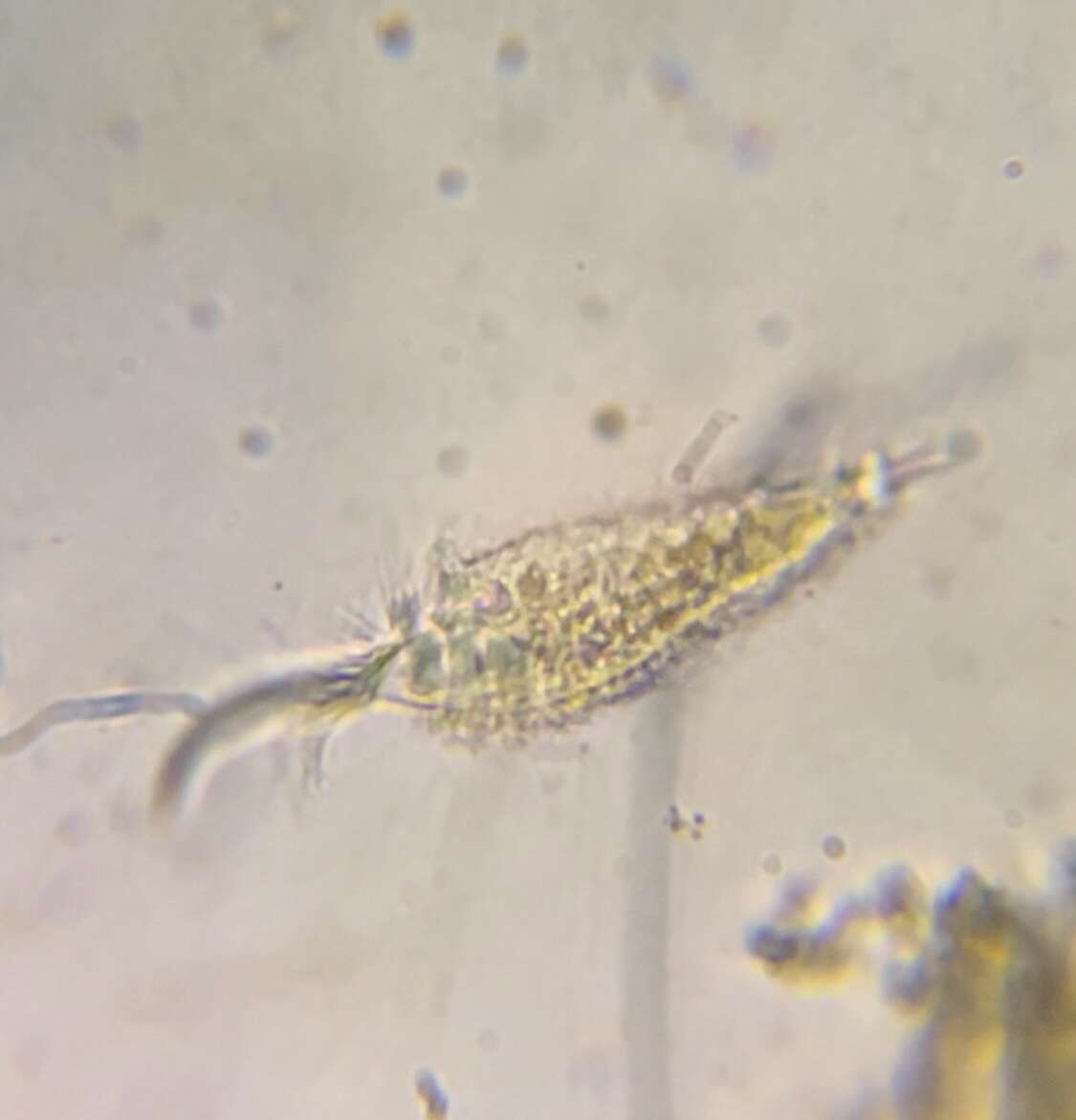
Portrait of Metopus striatus (synonyms include M. bacillatus, M. gibbus, M. pullus and M. acuminatus among others), a colorless metopid ciliate. The body is broadly domed anteriorly. The posterior end is bluntly tapered or may terminate in a short finger-like process. The oblique peristome with its adoral zone of membranelles terminates at the oral aperture in the posterior 1/3 of the body. The large spherical macronucleus is located in the midbody. There is one adjacent micronucleus. The sparse somatic kineties are longitudinal coursing to the left around the anterior dome (seen well here). There is a longer tuft of caudal cilia. The anterior refractile granules typical of metopids are much less conspicuous in this species. There is a prominent layer of subcortical extrusomes. A single contractile vacuole is located posteriorly. Collected from polysaprobic freshwater pond near Boise, Idaho September 2003. DIC optics.
-
Portrait of Metopus striatus (synonyms include M. bacillatus, M. gibbus, M. pullus and M. acuminatus among others), a colorless metopid ciliate. The body is broadly domed anteriorly. The posterior end is bluntly tapered or may terminate in a short finger-like process. The oblique peristome with its adoral zone of membranelles terminates at the oral aperture in the posterior 1/3 of the body. The large spherical macronucleus is located in the midbody. There is one adjacent micronucleus. The sparse somatic kineties are longitudinal coursing to the left around the anterior dome. There is a longer tuft of caudal cilia. The anterior refractile granules typical of metopids are much less conspicuous in this species. There is a prominent layer of subcortical extrusomes. A single contractile vacuole is located posteriorly. Collected from polysaprobic freshwater pond near Boise, Idaho September 2003. DIC optics.
-
-
-
-
-
-
-
-
-
-
-