-
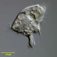
Portrait of the armophorid ciliate, Caenomorpha medusula (Perty,1852). The colorless pellicle is rigid and twisted to the left. The anterior is broadly rounded and umbrella-like with a long posterior spine. Somatic ciliature is reduced to 2 curved rows of thigmotactic cirri on the left side of the anterior dome (seen well here). The peristome spirals around the long axis, bordered anteriorly by a perizonal stripe of cilia and posteriorly by an adoral zone of membranelles. The cytostome situated at the posterior end of the peristome. There are 3 or 4 spherical macronuclei; 1 posterior contractile vacuole is located at the base of the spine . The cytoplasm contains multiple endosymbiotic methanogenic bacteria. C. medusula is found in anaerobic habitats. C. medusula is distinguished fro other species in the genus by its shape and by its two anterior cirral files (other species have only one). Collected from sapropelic bottom sediments of a slow-flowing freshwater stream near Boise, Idaho January 2005.Stained by the silver carbonate technic (see Foissner, W.Europ. J. Protistol.27,313-330;1991) Brightfield.
-
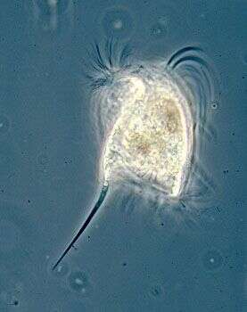
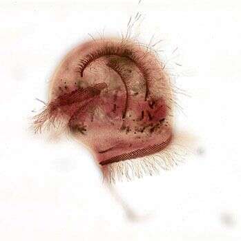
Phase contrast micrograph of living cell.
-
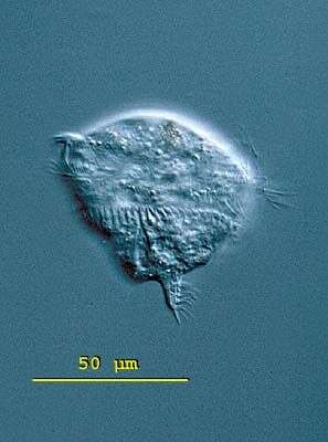
Caenomorpha (seen-owe-morph-a) uniserialis. Body medusoid with 1 to 3 posterior spines. Without somatic cilia except for a band which lies near the membranelles. The membranelles form a long spiral that encircles the body and ends in posteriorly situated cytostome. A contractile vacuole is located at the base of the spine. Symbiotic (methanogenic) bacteria occur in the cytoplasm. Move very quickly, with a jerky rotation. Between 1-4 macronuclei but always with only 1 micronucleus. Common in anoxic habitats. This slightly squashed specimen of Caenomorpha uniserialis was collected in a bog near Konstanz, Germany. The body of C. uniserialis is rather spherical. The short spine has a widened middle part. The cell is 72 microns long. Differental interference contrast.
-
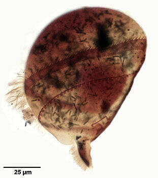
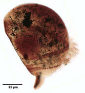
Silver carbonate preparation (see Foissner, W.Europ. J. Protistol.27,313-330;1991) of the sapropelic armophorid ciliate, Caenomorpha uniserialis (Levander, 1894), right lateral view. The cell body is parachute-shaped with a smooth broad anterior shield. The anterior shield bears a single file of cirri-like cilia (not visible in this view). The body terminates posteriorly in two spinous processes, a short simple one (not seen in this image) and a complex terminal longer spine with folds, projections and several rows of cilia (seen well here). The perizonal ciliary stripe originates on the left lateral aspect anteriorly and makes a complete turn around the body along the spiraling peristome and terminates on the same longitudinal line posteriorly (seen here). The area of the cytostome is between the two spinous processes at the posterior end of the peristome. An adoral zone of membranelles posterior to the perizonal stripe of kineties parallels the spiral peristome (seen well here). An undulating membrane originates near the posterior end of the perizonal stripe between it and the adoral zone of membranelles and terminates at the cytostome (seen as a thin dark line in this image). The cytoplasm contains several forms of endosymbiotic bacilli (stained dark black in this image). The contractile vacuole (not seen here) is located at the base of the large spine. C. uniserialis is bactiverous. Collected from stagnant freshwater bottom sediment with rotting leaves and rich in hydrogen sulfide near Boise, Idaho August 2004. Brightfield optics.
-

Silver carbonate preparation (see Foissner, W.Europ. J. Protistol.27, 313-330; 1991) of the sapropelic armophorid ciliate, Caenomorpha uniserialis (Levander, 1894), left lateral view. The cell body is parachute-shaped with a smooth broad anterior shield. The anterior shield bears a single file of cirri-like cilia (seen well here). The body terminates posteriorly in two spinous processes, a short simple one (not seen in this image) and a complex terminal longer spine with folds, projections and several rows of cilia. The perizonal ciliary stripe originates on the left lateral aspect anteriorly and makes a complete turn around the body along the spiraling peristome and terminates on the same longitudinal line posteriorly (seen well here). The area of the cytostome is between the two spinous processes at the posterior end of the peristome. An adoral zone of membranelles parallels the spiral peristome (seen well here). An undulating membrane originates near the posterior end of the perizonal stripe and terminates at the cytostome (seen as a thin dark line in this image). The single large spherical macronucleus and the overlying micronucleus are seen well here. The cytoplasm contains several forms of endosymbiotic bacilli (stained dark black in this image). The contractile vacuole (not seen here) is located at the base of the large spine. C. uniserialis is bactiverous. Collected from stagnant freshwater bottom sediment with rotting leaves and rich in hydrogen sulfide near Boise, Idaho August 2004. Brightfield optics.
-
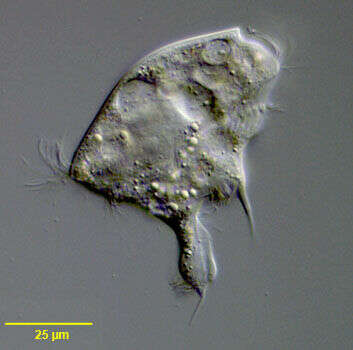
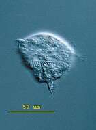
Portrait of the sapropelic armophorid ciliate, Caenomorpha uniserialis. The cell body is parachute-shaped with a smooth broad anterior shield. The anterior shield bears a single file of cirri (not seen here). The body terminates posteriorly in two spinous processes, a short simple one and a complex longer spine with folds and projections. The perizonal ciliary stripe winds along the spiraling peristome and terminates on the large posterior spine (seen well here). The area of the cytostome is seen between the two spinous processes at the posterior end of the peristome. An adoral zone of membranelles parallels the spiral peristome. The single large spherical macronucleus and the overlying micronucleus are seen well here. The yellowish refractile droplets in the cytoplasm may represent lipid. Many food vacuoles are present. The cytoplasm contains several forms of endosymbiotic bacteria. The contractile vacuole (not seen here) is located at the base of the large spine. C. uniserialis is bactiverous. Collected from stagnant freshwater bottom sediment with rotting leaves and rich in hydrogen sulfide near Boise, Idaho January 2004. DIC optics