-
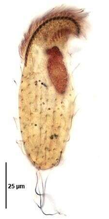
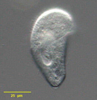
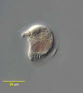
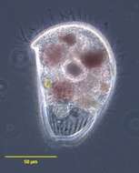
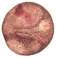
Ventral view of the infraciliature of Metopus hasei (SONDHEIM,1929).Only the anterior portion of the adoral zone of membranelles is well impregnated in this image. The dark band bordering the peristome is the perizonal ciliary stripe consisting of five rows of dikinetids.The tuft of long caudal cilia is seen here.From a rewetted soil sample collected from the margin of a eutrophic freshwater pond near Boise, Idaho. Stained by the silver carbonate technique (see Foissner, W.Europ. J. Protistol.27:313-330;1991).Brightfield.
-
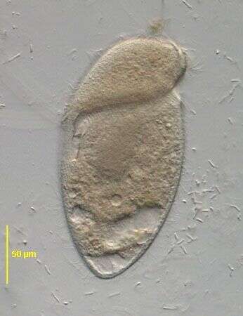
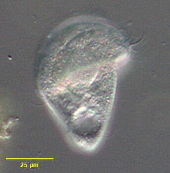
Portrait (anterior oblique view) of the metopid ciliate, Brachonella galeata (Kahl 1927 [Metopus galeatus]; Jankowski, 1964). The anterior half of the cell is broadly helmet-shaped. The posterior is bluntly conical. The peristome encircles the body at the junction of the anterior and posterior halves of the cell, the origin lying just anterior to the termination on the same longitudinal line. The peristome terminates in a posterior cytostome. There is an adoral zone of membranelles on the left margin of the peristome. The posterior part of the AZM is seen in this image. The somatic cilia are long and sparse. There is a tuft of longer cilia at the posterior end. A single ellipsoid macronucleus is located centrally or anteriorly (not seen well here). There is a single irregularly shaped contractile vacuole at the posterior end. There are purple sulfur bacteria visible in food vacuoles. B. galeata is anaerobic. Collected from anoxic sediment of slow-moving freshwater stream near Boise, Idaho in April 2004. DIC optics.
-
Caenomorpha simplex (Jankowski,1964).Phase contrast.
-
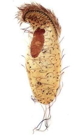
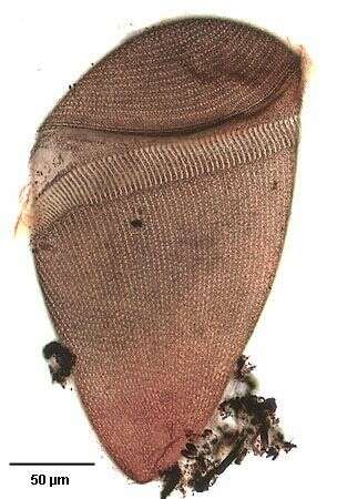
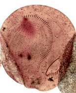
Dorsal view of the infraciliature of Metopus hasei (SONDHEIM,1929).The tuft of long caudal cilia is seen here.From a rewetted soil sample collected from the margin of a eutrophic freshwater pond near Boise, Idaho. Stained by the silver carbonate technique (see Foissner, W.Europ. J. Protistol.27:313-330;1991).Brightfield.
-
Portrait (coronal section) of the metopid ciliate, Brachonella galeata (Kahl 1927 [Metopus galeatus]; Jankowski, 1964). The anterior half of the cell is broadly helmet-shaped. The posterior is bluntly conical. The peristome encircles the body at the junction of the anterior and posterior halves of the cell, the origin lying just anterior to the termination on the same longitudinal line. The peristome terminates in a posterior cytostome. There is an adoral zone of membranelles on the left margin of the peristome. The somatic cilia are long and sparse. There is a tuft of longer cilia at the posterior end. A single ellipsoid macronucleus is located centrally or anteriorly . There is a single irregularly shaped contractile vacuole at the posterior end. There are purple sulfur bacteria visible in food vacuoles. B. galeata is anaerobic. Collected from anoxic sediment of slow-moving freshwater stream near Boise, Idaho in April 2004. DIC optics.
-
Metopus fuscus (Kahl,1927).Brightfield, condenser closed.
-
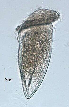
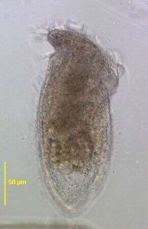
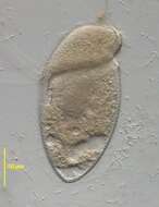
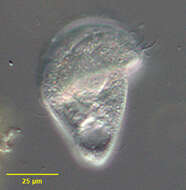
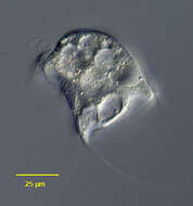
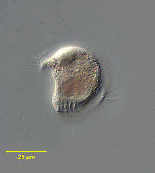
Portrait of Metopus spinosus a polysaprobic ciliate (ventral view). Synonymous with Metopus caudatus. The body is elongate and slightly flattened dorsoventrally. The rounded anterior is twisted to the left and the posterior is drawn out to a tapered point. There is an anterior apical aggregate of refractile granules typical of metopids. Somatic kineties are longitudinal and uniform. The peristome slants obliquely across the ventral surface. There is a prominent adoral zone of membranelles on its left and several kineties of longer cilia along its right margin. The cytostome is at the right posterior end of the peristome. The large ellipsoid central macronucleus is not seen in this image. There is a posterior contractile vacuole.
-
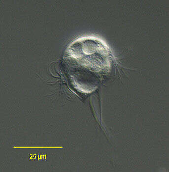
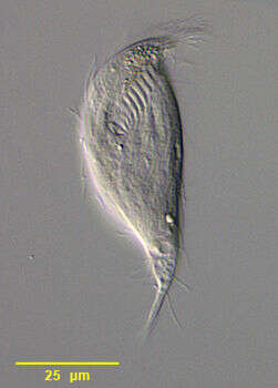
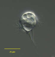
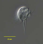
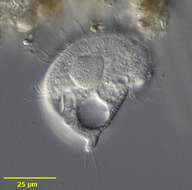
Portrait of the small caenomorphid ciliate, Ludio parvulus (Penard, 1922). The genus is monotypic. The colorless cell is roughly pyriform. The posterior end terminates in a short conical spinous projection. The pellicle is firm and only slightly deformable. The subequatorial peristome spirals around the body. The peristome is bordered anteriorly by a narrow perizonal ciliary stripe and also by an inconspicuous adoral zone of membranelles. The cytostome is at the posterior end of the peristome. A needle-like spine at least as long as the cell body arises on the right surface and extends posteriorly. A prominent cirrus approximately the same length as this spine trails behind the cell. A shorter anterior cirrus is not well seen in this image. The single spherical macronucleus is located eccentrically. The micronucleus is not clearly seen in these images. There is a single posterior contractile vacuole. L. parvulus is sapropelic. L. parvulus lacks the anterior rows of cirri seen in both Caenomorpha and Cirranter. Unlike Ludio, Cirranter lacks spines. Collected from a slow-moving freshwater stream near Boise, Idaho December 2004. DIC.
-
Metopus fuscus (Kahl,1927). DIC.
-
Metopus violaceus (KAHL, 1927). Phase contrast.
-
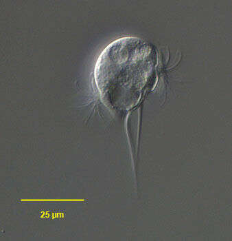
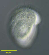
Portrait of the small caenomorphid ciliate, Ludio parvulus (Penard, 1922). The genus is monotypic. The colorless cell is roughly pyriform. The posterior end terminates in a short conical spinous projection. The pellicle is firm and only slightly deformable. The subequatorial peristome spirals around the body. The peristome is bordered anteriorly by a narrow perizonal ciliary stripe and also by an inconspicuous adoral zone of membranelles. The cytostome is at the posterior end of the peristome. A needle-like spine at least as long as the cell body arises on the right surface and extends posteriorly. A prominent cirrus approximately the same length as this spine trails behind the cell (seen here to viewr's left of long posterior spine). A shorter anterior cirrus is not well seen in this image. The single spherical macronucleus is located eccentrically. The micronucleus is not clearly seen in these images. There is a single posterior contractile vacuole. L. parvulus is sapropelic. L. parvulus lacks the anterior rows of cirri seen in both Caenomorpha and Cirranter. Unlike Ludio, Cirranter lacks spines. Collected from a slow-moving freshwater stream near Boise, Idaho December 2004. DIC.
-
Metopus fuscus (Kahl, 1927). Brightfield, condenser closed.
-
Metopus violaceus (KAHL, 1927). DIC.
-
Metopus fuscus (Kahl,1927). Stained by the silver carbonate technique (see Foissner, W. Europ. J. Protistol., 27:313-330;1991).Brightfield.
-
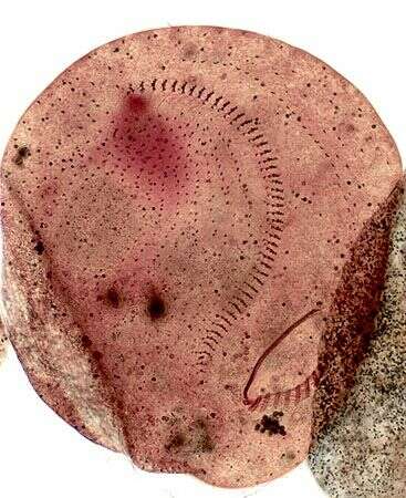
Infraciliature of Metopus violaceus (KAHL, 1927). Stained by the silver carbonate technique (see Foissner, W. Europ. J. Protistol., 27:313-330;1991).Brightfield
-
Infraciliature of Metopus violaceus (KAHL, 1927). Stained by the silver carbonate technique (see Foissner, W. Europ. J. Protistol., 27:313-330;1991). Brightfield.
-
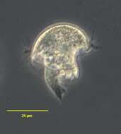
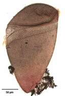
Ventral view of Metopus striatus (synonyms include M. bacillatus, M. gibbus, M. pullus and M. acuminatus among others), a colorless metopid ciliate. The body is broadly domed anteriorly. The posterior end is bluntly tapered or may terminate in a short finger-like process. The oblique peristome with its adoral zone of membranelles terminates at the oral aperture in the posterior 1/3 of the body (seen well here). The large spherical macronucleus is located in the midbody. There is one adjacent micronucleus. The sparse somatic kineties are longitudinal coursing to the left around the anterior dome. There is a longer tuft of caudal cilia. The anterior refractile granules typical of metopids are much less conspicuous in this species. There is a prominent layer of subcortical extrusomes. A single contractile vacuole is located posteriorly. Collected from polysaprobic freshwater pond near Boise, Idaho September 2003. DIC optics.
-
Optical section through Metopus striatus (synonyms include M. bacillatus, M. gibbus, M. pullus and M. acuminatus among others), a colorless metopid ciliate. The body is broadly domed anteriorly. The posterior end is bluntly tapered or may terminate in a short finger-like process. The oblique peristome with its adoral zone of membranelles terminates at the oral aperture in the posterior 1/3 of the body. The large spherical macronucleus is located in the midbody. There is one adjacent micronucleus. The sparse somatic kineties are longitudinal coursing to the left around the anterior dome. There is a longer tuft of caudal cilia. The anterior refractile granules typical of metopids are much less conspicuous in this species. There is a prominent layer of subcortical extrusomes. A single contractile vacuole is located posteriorly. Collected from polysaprobic freshwater pond near Boise, Idaho September 2003. DIC optics.
-
Portrait of Metopus striatus (synonyms include M. bacillatus, M. gibbus, M. pullus and M. acuminatus among others), a colorless metopid ciliate. The body is broadly domed anteriorly. The posterior end is bluntly tapered or may terminate in a short finger-like process. The oblique peristome with its adoral zone of membranelles terminates at the oral aperture in the posterior 1/3 of the body. The large spherical macronucleus is located in the midbody. There is one adjacent micronucleus. The sparse somatic kineties are longitudinal coursing to the left around the anterior dome (seen well here). There is a longer tuft of caudal cilia. The anterior refractile granules typical of metopids are much less conspicuous in this species. There is a prominent layer of subcortical extrusomes. A single contractile vacuole is located posteriorly. Collected from polysaprobic freshwater pond near Boise, Idaho September 2003. DIC optics.
-
Portrait of Metopus striatus (synonyms include M. bacillatus, M. gibbus, M. pullus and M. acuminatus among others), a colorless metopid ciliate. The body is broadly domed anteriorly. The posterior end is bluntly tapered or may terminate in a short finger-like process. The oblique peristome with its adoral zone of membranelles terminates at the oral aperture in the posterior 1/3 of the body. The large spherical macronucleus is located in the midbody. There is one adjacent micronucleus. The sparse somatic kineties are longitudinal coursing to the left around the anterior dome. There is a longer tuft of caudal cilia. The anterior refractile granules typical of metopids are much less conspicuous in this species. There is a prominent layer of subcortical extrusomes. A single contractile vacuole is located posteriorly. Collected from polysaprobic freshwater pond near Boise, Idaho September 2003. DIC optics.
-
-
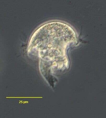
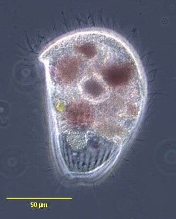
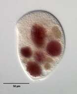
Caenomorpha (seen-owe-morph-a) medusula. Body medusoid with 1 to 3 posterior spines. Without somatic cilia except for a band which lies near the membranelles. The membranelles form a long spiral that encircles the body and ends in posteriorly situated cytostome. A contractile vacuole is located at the base of the spine. Symbiotic (methanogenic) bacteria occur in the cytoplasm. Move very quickly, with a jerky rotation. Between 1-4 macronuclei but always with only 1 micronucleus. Common in anoxic habitats. This specimen was collected in a bog near Konstanz, Germany. Slightly squashed specimen of Caenomorpha medusula. Focal plane on the rim of the bell-shaped anterior half of the body. Specimen measures 90 microns. Differential interference contrast.
-
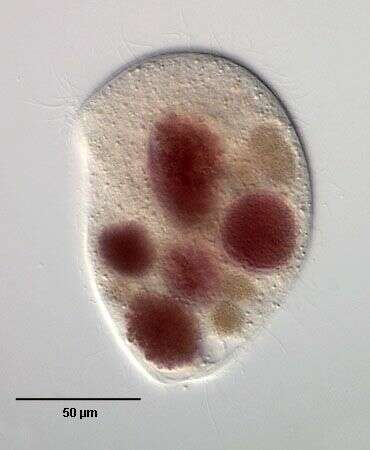
Caenomorpha (seen-owe-morph-a) medusula. Body medusoid with 1 to 3 posterior spines. Without somatic cilia except for a band which lies near the membranelles. The membranelles form a long spiral that encircles the body and ends in posteriorly situated cytostome. A contractile vacuole is located at the base of the spine. Symbiotic (methanogenic) bacteria occur in the cytoplasm. Move very quickly, with a jerky rotation. Between 1-4 macronuclei but always with only 1 micronucleus. Common in anoxic habitats. This specimen was collected in a bog near Konstanz, Germany. The two macronuclei are visible near the center of the body and the contractile vacuole is located at the base of the spine. 90 microns long. Differential interference contrast.
-
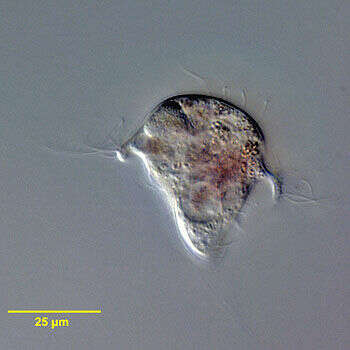
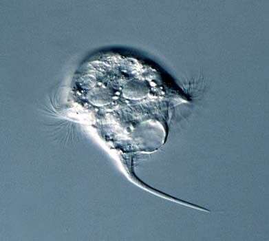
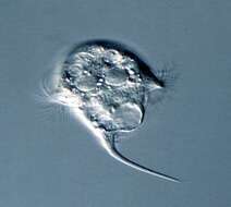
Portrait of the armophorid ciliate, Caenomorpha medusula (Perty,1852). The colorless pellicle is rigid and twisted to the left. The anterior is broadly rounded and umbrella-like with a long posterior spine. Somatic ciliature is reduced to 2 rows of thigmotactic cirri on the left side of the anterior dome. The peristome spirals around the long axis, bordered anteriorly by a perizonal stripe of cilia and posteriorly by an adoral zone of membranelles. The cytostome situated at the posterior end of the peristome. There are 3 or 4 spherical macronuclei; 1 posterior contractile vacuole is located at the base of the spine . The cytoplasm contains multiple endosymbiotic methanogenic bacteria. C. medusula is found in anaerobic habitats. C. medusula is distinguished fro other species in the genus by its shape and by its two anterior cirral files (other species have only one). Collected from sapropelic bottom sediments of a slow-flowing freshwater stream near Boise, Idaho January 2005. DIC.