“Obscuranella papyrodes new species (Figures 1-36, Table 1)
Bathydomus obtectus Thiele, 1912—Dell, 1990:198-199, figs. 299-300.
Bathydomus sp.—Dell, 1990:199.
Table 1. Obscuranella papyrodes, new species. Measurements of shell characters. Linear measurements in mm.
Character
Holotype
Paratype 1
Paratype 3
USNM 901317
USNM 870610
USNM 870610
Shell Length (SL)
63+
58+
33+
57.0
43.6+
33.2+
Last Whorl Length (LWL)
55.2
51.8
28.5
47.3
38.5
30.4
Aperture Length (AL)
45.1
43.3
23.0
38.4
31.5
25.5
Shell Width (SW)
40.8
41.5
19+
34.1
24+
20.2+
Number of spiral cords on last whorl
14.
12
14
5
14
13
Number of spiral cords on penultimate whorl
6
7
7
2
5
3
Description: Shell large (exceeding 63 mm), very thin, fragile, ovate-pyriform. Protoconch and upper whorls missing in all type material. Preserved portions of teleoconch of 2 ½ rapidly expanding, evenly rounded whorls. Shoulder rounded, indistinct. Suture adpressed, shallow. Axial sculpture limited to fine, straight, weakly prosocline growth lines. Adult specimens with a single, weak, hollow varix adjacent to thin, flared outer lip (figures 2, 7, arrow). Spiral sculpture of sharp, narrow, evenly spaced cords (14 on last whorl, 6 on penultimate whorl), with much weaker sinuous threads (22-30) of varying width between adjacent cords. Aperture large [~0.7 shell length (SL)], broadly ovate, deflected from shell axis by 9-11°. Outer lip thin, evenly rounded in upper part and concave at transition to siphonal canal, weakly reflected. Inner lip consisting of long, convex, medially indented parietal region and short, smooth, axial columella with strong, long siphonal fold that crosses coiling axis of shell. Siphonal canal short, broad, weakly recurved dorsally. Callus of thin, white, porcellaneous glaze overlying parietal region, adapical portion of broad, nearly axial siphonal fasciole. Shell color pale olive-tan, confined to outermost shell layer. Aperture white. Periostracum very thin, yellowish brown, with densely spaced axial lamellae, occasional short hairs at intersection of lamellae with spiral cords. Operculum (figures 4, 5) very small (0.16 AL), vestigial, dark yellow, subtriangular, with straight sides, terminal nucleus. Dorsal surface with numerous, closely spaced growth lines. Ventral surface with thin, glazed lateral margins. Operculum attached over most of its surface.
Shell ultrastructure (Figure 23): Shell thin (101 μm), composed of three layers. Outermost layer (figure 23, ca) thinnest (4 μm), composed of columnar crystals. Middle layer (figure 23, ccl) thickest (79 µm), composed of collabrally oriented cross-lamellar crystals. Inner layer (figure 23, rcl) thin (18 µm), composed of cross-lamellar crystals oriented perpendicular to growing edge of the shell.
Anatomy (Paratype 1, ♀): Soft tissues comprising approximately 3 ½ whorls. Mantle cavity spans just under ½ whorl, nephridium (figures 24, 25, 27, n) about ¼ whorl, digestive gland (figures 24, 25, dg) 2 ½ whorls. Mantle edge (figures 24, 25, 27, me) thickened, smooth, completely covers head. Columellar muscle (figure 25, cm) thick, broad, spanning slightly more than one whorl, attached to shell at rear of nephridium. Foot short in contracted state (Length/Width ≈ 1.0), with conspicuous propodium. Body color uniform reddish-tan, without pattern in alcohol preserved specimens. Head (figure 26) very large, as wide as foot, with broad, blunt, tapering tentacles (figure 26, tn) with black eyes at their bases. Operculum about 4 mm long (0.09 AL), otherwise similar to that of holotype. Paratype 1 (and all other preserved specimens) with proboscis protruded through very wide rhynchostome (figure 26).
Mantle cavity (Figure 27): Mantle cavity as deep as broad (~ ½ whorl). Siphon (figures 24, 25, 27, s) broad, muscular, very short, extending slightly beyond mantle edge (figures 24-27, me). Osphradium (figures 24-27, os) situated along central half of ctenidium, bipectinate, nearly symmetrical, slightly narrower on left side than right. Ctenidium (figures 24-27, ct) long, spanning nearly entire mantle length, formed of tall triangular lamellae, nearly twice as high as broad. Hypobranchial gland (figure 27, hg) poorly developed, lacking distinct folds. Rectum (figure 27, re) runs along inner surface of pallial oviduct, narrow, terminating in simple anus (figure 27, a) behind thickened mantle edge.
Alimentary system (Figures 15, 19-22, 24, 32-35): Everted proboscis (figures 24-26, pr) ~30 mm long (0.7 AL), unpigmented, with folded walls indicating potential for further extension. Proboscis wall very thick, ~60% of proboscis radius, composed of 3 layers of muscles. Innermost laver of circular muscles (figure 32, cm), middle layer thickest (2/3 of proboscis wall), of longitudinal muscles (figure 32, lm), outer layer of circular muscle. Buccal mass (figure 33, bm) small, attached to proboscis walls by numerous, thin tensor muscles (figures 15, 32, 33, tm), as is the anterior oesophagus (figures 15, 32, aoe). Retractor muscles passing through nerve ring and joining buccal mass and columellar muscle absent. Mouth figure 32, mo) a narrow, vertical slit. Buccal tube figure 33, bt) short, leading to cuticle-lined buccal cavity with ventral pair of semicircular jaws (figure 33, j). Jaws figures 16-18) dark brown, pappilate along outer edge figure 17). Inner surface of jaw composed of small, closely spaced platelets that produce "cobbled" surface distally (figure 18), smooth proximally (figure 16). Odontophore (figure 15, od) small, oval, lining bottom of the buccal cavity. Walls of buccal cavity very thick. Proboscis nerves (figure 32, pn) paired, very thick, running from cerebro-pleural ganglia along proboscis length, innervating buccal mass and anterior part of proboscis. Anterior esophagus divided into dorsal and ventral channels by prominent longitudinal folds (figures 15, 34, 35, lf) that extend from the buccal cavity to the posterior edge of esophageal gland. The right fold overlaps the left (figure 34). Radular ribbon (figure 19) short (5.8 mm, 0.13 AL), nearly twice as long as cartilages, narrow (~580 µm, 0.013 AL), consisting of 45 rows of teeth, posteriormost 4 rows nascent. Rachidian tooth (figure 21, rt) with large, broad median cusp, flanked by 5-9 denticles per side. Base broad, strongly concave posteriorly, lacking cusps along tooth base. Lateral teeth (figures 21, 22, lt) narrow, with long, thin, cusp flanked by 3-4 denticles on inner edge, 4-6 denticles on outer edge. Two long, recurved, distally flattened marginal teeth (figures 20; 22, mt) per side, outer tooth longer than inner. Inner distal edges serrated with 2-7 cusps. Salivary glands large, irregularly shaped, completely covering the esophageal gland (figure 32, oeg). Right salivary gland more elongated, slightly larger than left. Each gland consists of two lobes. Posterior lobe (figure 32, plsg) massive, composed of curved radially oriented blind tubules. Anterior lobe (figure 32, alsg) smaller, acinous, ventral. Salivary ducts (figures 32‑34, sd) thick, extending from posterior lobes, becoming attached to oesophagus walls before passing through nerve ring. We were not able to identify connections between the salivary ducts and the anterior lobes of the salivary glands, as reported for Cymatium intermedium (Pease, 1869) by Andrews et al. (1999). Salivary glands attached to oesophagus by thin muscular and connective tissue fibers and innervated by several nerve branches. Esophageal gland formed of deeply glandular dorso-ventral folds, small relative to salivary glands, of the same color as surrounding tissues and esophagus. Stomach large, U-shaped, similar to that of Cymatium nicobaricum (Houbrick and Fretter, 1969) in external morphology, too poorly preserved to reveal details of internal morphology, except that the ducts of the digestive gland are paired, closely spaced, and situated in the middle region of the stomach. Posterior duct twice the diameter of anterior duct. After leaving stomach intestine curves dorsally, passing under then along posterior edge of nephridium towards rear of mantle cavity (figures 24, 25, in).
Female reproductive system (Figures 25, 27-30): The only specimen dissected (Paratype 1) was a mature female. Pallial oviduct consists of small albumen gland (figure 25, ag) along outer wall of nephridium, large capsule gland (figures 25, 27, cg) lining right wall of pallial cavity. Lumen of capsule gland small anteriorly (figure 29), broad posteriorly (figure 28), at juncture to albumen gland. No seminal receptacles were identified, possibly due to poor fixation. Bursa copulatrix (figures 27, 29, 30, bc) large, long, spanning more than half length of capsule gland. Pallial oviduct opens (figures 27, 29, go) by long, narrow slit at midlength.
Male reproductive system (Figure 31): The reproductive system of a male specimen lacking shell (paratype 2) was examined. Testes occupy upper half of whorls of visceral mass above posterior border of stomach, giving rise to seminal duct. Seminal duct forms large, convoluted seminal vesicle before entering mantle cavity. Duct descends to floor of mantle cavity at mid-length, opening to form groove with thickened, obviously glandular walls leading to penis base. Penis (figure 31, p) broad, dorsoventrally flattened, with seminal groove (figure 31, sgr) running along inner edge, around the bluntly rounded distal edge, halfway down the outer edge, terminating in a very small, inconspicuous papilla (figure 31, pap).
Type locality: Victoria Land, Balleny Islands, Sturge Island, R/V Eltanin, st. 1949, 66°52' S, 164°32'E, in 2507-2525 m, 5 February 1967.
Type material: Holotype, USNM 898683 (figures 1-5, shell and operculum only, soft parts not present), from the type locality; paratype 1, USNM 896131 (figures 6–7, ♀pecimen, anatomical descriptions based on this specimen), paratype 2, USNM 896139 (♂ specimen, shell not present, male reproductive system based on this specimen), both from south of Hjort Seamount, R/V Eltanin stn. 1964, 59°58'S, 155°31'W, in 2985-2992 m, 10 February 1967: paratype 3, USNM 886105 (figures 11-12, sex undetermined) Victoria Land, south of Scott Island Bank. R/V Eltanin, stn. 1939, 69°03' S, 179°41’ E in 3519-3596 m, 1 February 1967.
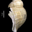
Other material examined: USNM 901317 (Figs 13-14, 1 specimen, sex undetermined) Victoria Land, Moubray Bay, R/V Eltanin, stn. 2002, 72°18'S, 177°35'E, in 2005-2010 m, 11 January 1968; USNM 870610 (figures 8-10, 2 dead shells) Antarctic Peninsula, R/V Eltanin, stn. 1003, 62°41'S, 54°43'W, in 210-220 m, 15 March 1964.
Etymology: papyrodes—made from papyrus, referring to the thinness of the shell.
Remarks: The type series of Obscuranella papyrodes, n. sp., consists of four specimens, including one paratype that lacks a shell. All were collected living on the abyssal plain off the Ross Sea. We are provisionally attributing three additional specimens to this species. One live-collected specimen (figure 13; USNM 901317), also from the abyssal plain off the Ross Sea, was considered by Dell (1990:199) to be congeneric but not conspecific with Obscuranella papyrodes n. sp. (which he identified as Bathydomus obtectus Thiele, 1912) because of its more elongated shell, longer siphonal canal, and angular shoulder. Even the earliest teleoconch whorls of this specimen appear angular because of a prevalent spiral cord along the periphery. This is exaggerated by a break in the shell and subsequent repair, evidenced by a thickened scar on the posterior part of the last whorl. The elongated shell and prominent siphonal canal are features shared with an immature paratype (paratype 3, figures 11-12) of 0. papyrodes. This is the only specimen of Obscuranella with some portion of the early whorls intact (figure 14). The protoconch (2.0 mm estimated diameter) is eroded and replaced by a plug, but the earliest teleoconch whorls are well preserved and clearly show spiral cords.
Two dead collected shells (figures 8-10; USNM 870610), labeled as coming from upper slope depths (210-220 m) off the Antarctic Peninsula, closely match the morphology of this new species. We regard these specimens to represent 0. papyrodes, but are skeptical of the accuracy of the locality data. Not only is this location on the opposite side of the Antarctic continent from all records of live collected 0. papyrodes, it is also from much shallower depths (220 m vs. 2000+ m).
DISCUSSION
Obscuranella can readily be attributed to the superfamily Tonnoidea on the basis of its pyriform shell with large aperture and conspicuous, if short siphonal canal; its extensible proboscis; its taenioglossan radula; its large salivary glands composed of morphologically differentiated anterior and posterior lobes and salivary ducts that pass through the nerve ring, as well as its undifferentiated oesophageal gland. It can be excluded from Ficidae, which was removed from Tonnoidea and elevated to superfamily status by Riedel (1994), by its high spire, lack of long siphonal canal, and also because Ficidae is characterized by small, tubular salivary glands. Similarly, it can be excluded from Laubierinidae, a family diagnosed by its monopectinate osphradium and excluded from Tonnoidea by Bandel and Riedel (1994), by its nearly symmetrical, bipectinate osphradium.
The shell of Obscuranella suggests an affinity with the deep-sea family Pisanianuridae (originally proposed as a subfamily of Ranellidae by Warén and Bouchet, 1990, transferred to Laubierinidae by Bandel and Riedel, 1994, and elevated to family status by Beu, 1998) by virtue of its smooth shell lacking regular varices and weakly defined anterior canal. The operculum of Pisanianura is slightly coiled but has a terminal nucleus, as does Obscuranella. However, the rachidian teeth of Obscuranella lack the lateral basal denticles present in Pisanianuridae (e.g. Warén and Bouchet, 1990:figs. 25-27), Bursidae (e.g. Warén and Bouchet, 1990:figs. 6, 8), Tonnidae (e.g. Warén and Bouchet, 1990:figs. 9-14), and Laubierinidae (e.g. Warén and Bouchet, 1990:figs. 4144), but absent in Cassidae (e.g. Warén and Bouchet, 1990:figs. 15, 16, 18), Personidae (e.g. Beu, 1998:fig. 15.140 E), and Ranellidae (e.g. Warén and Bouchet, 1990:figs. 28, 30, 32, 40).
The shell of Obscuranella somewhat resembles that of Oocorys sulcata Fischer, 1883 (Oocorythinae, Cassidae) (see, eg. Bouchet and Warén, 1993:figs. 1936-1943), and some ranellids, such as Argobuccinum pustulosum (Lightfoot, 1786) (see e.g. Beu, 1998:fig. 15.12 D). The operculum of Obscuranella has a sharply pointed, terminal nucleus, and differs from the spirally coiled operculum of Oocorys (Warén and Bouchet, 1990: fig. 66). In adult Argobuccinum the nucleus of the operculum is subcentral, but in very young specimens of A. pustulosum (Warén and Bouchet, 1990:fig. 69) the nucleus is terminal. However, the operculum of Obscuranella is distinctive in its very small size, relative to the aperture, and in having straight, anteriorly converging margins.
The anatomy of Obscuranella is typically tonnoidean, most closely resembling that of the ranellid Cymatium (Houbrick and Fretter, 1969). Obscuranella can be distinguished anatomically from the Tonnidae by its lack of a buccal gland, and from Pisanianuridae and Laubierinidae by its lack of long proboscis retractor muscles that pass through the nerve ring.
We assign this genus to the family Ranellidae because of it general similarity to Argobuccinum in shell form, radular morphology, and gross anatomy. Moreover, Ranellidae is the only tonnoidean family to occur in Antarctic waters—Fusitriton magellanicus (Röding, 1798), a species with a wide geographic range, has been taken from several stations in the Weddell and Ross Quadrants (e.g. USNM 896058, USNM 896103. USNM 896277, USNM 898520—see Polar Invertebrate Catalog http:// www.nmnh.si.edu/cgi-bin/wbd/iz/pci/form). Obscuranella papyrodes represents the first record of Ranellidae from abyssal depths."
(Kantor & Harasewych: 2000, 103-111)