-
All Biocode files are based on field identifications to the best of the researcher’s ability at the time.
-
Fei Dong, Chungkun Shih, Dong Ren
Zookeys
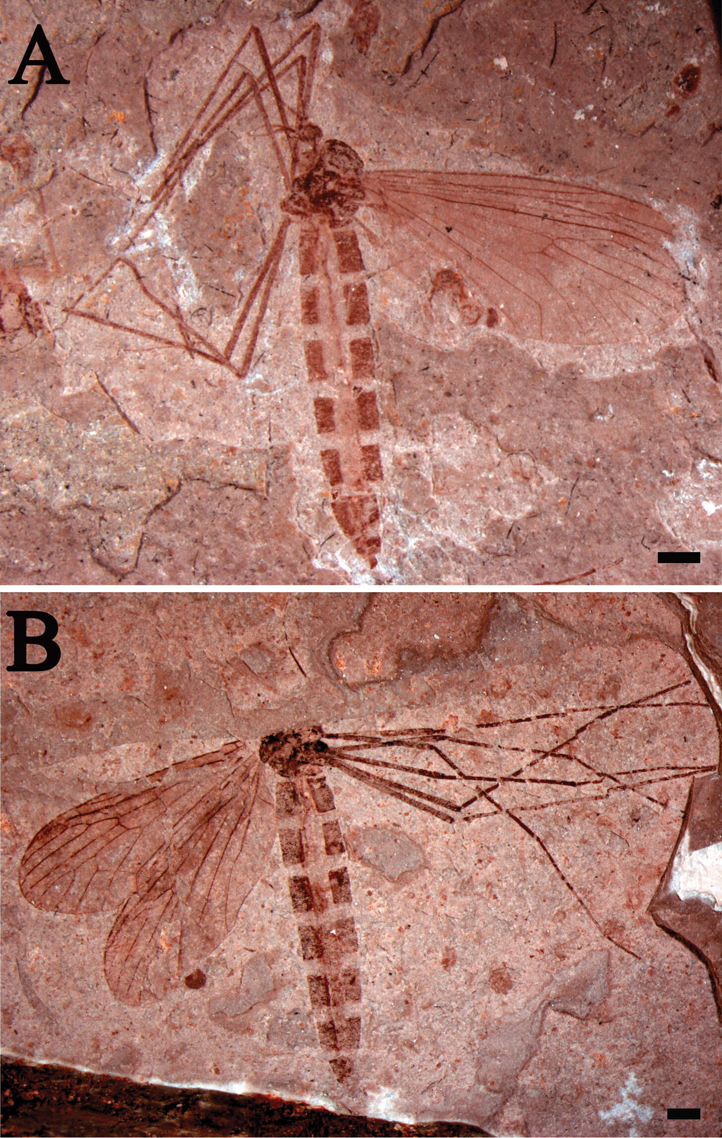
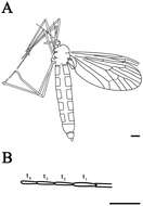
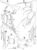
Figure 1.Eotrichocera (Archaeotrichocera) longensis sp. n. Holotype, specimen CNU-DIP-NN2013133 A Photograph. Paratype, specimen CNU-DIP-NN2013131 B Photograph. Scale bars = 1 mm.
-
All Biocode files are based on field identifications to the best of the researcher’s ability at the time.
-
Fei Dong, Chungkun Shih, Dong Ren
Zookeys
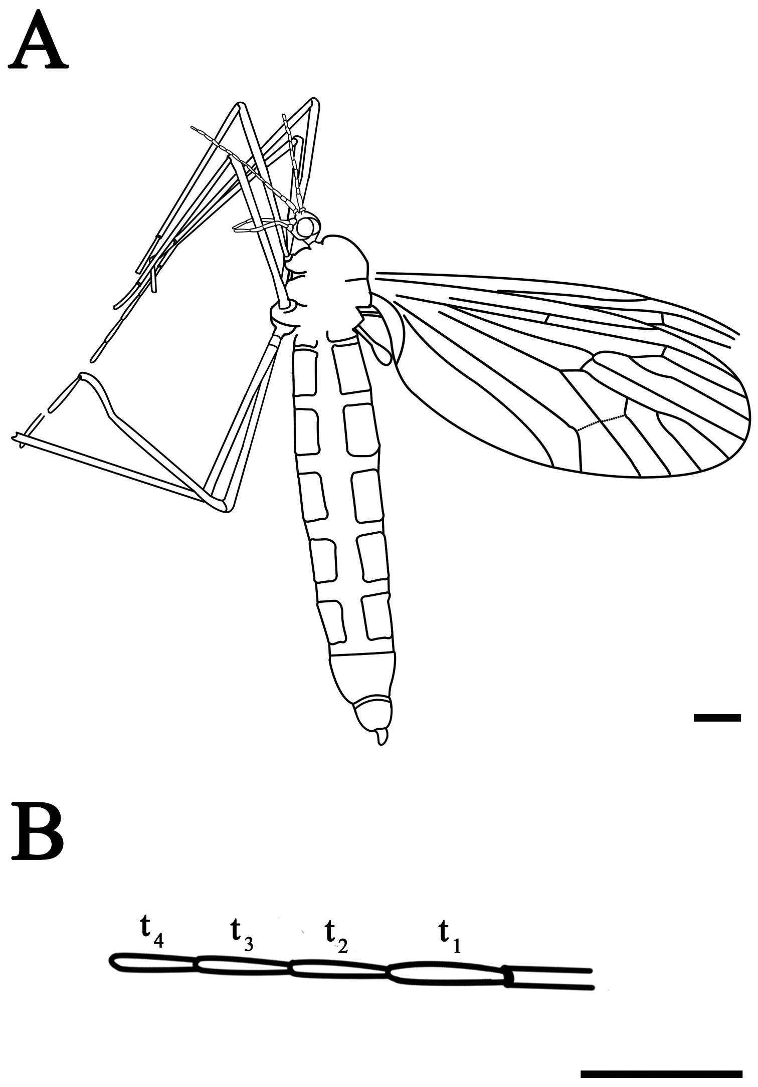
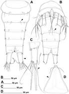
Figure 2.Eotrichocera (Archaeotrichocera) longensis sp. n. Holotype, specimen CNU-DIP-NN2013133 A Line drawing B Tarsus of the mid leg. Scale bars = 1 mm; t1 = the first segment of tarsus; t2 = the second segment of tarsus; t3 = the third segment of tarsus; t4 = the fourth segment of tarsus.
-
Fei Dong, Chungkun Shih, Dong Ren
Zookeys
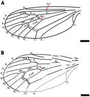
Figure 3.Eotrichocera (Archaeotrichocera) longensis sp. n. Holotype, specimen CNU-DIP-NN2013133 A Line drawing of left wing. Paratype, specimen CNU-DIP-NN2013131 B Line drawing of left wing. Scale bars = 1 mm
-
Tomislav Karanovic, Kichoon Kim, Wonchoel Lee
Zookeys
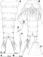
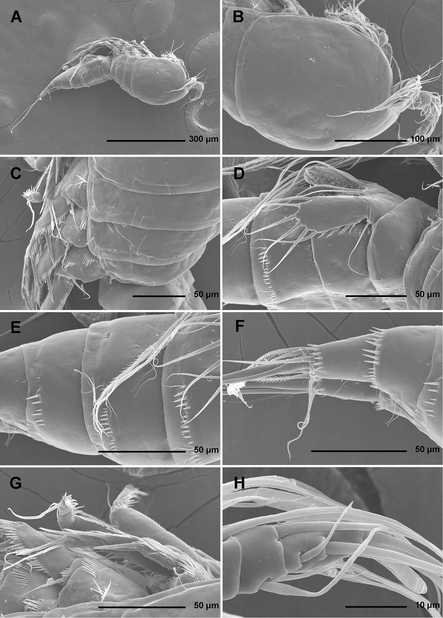
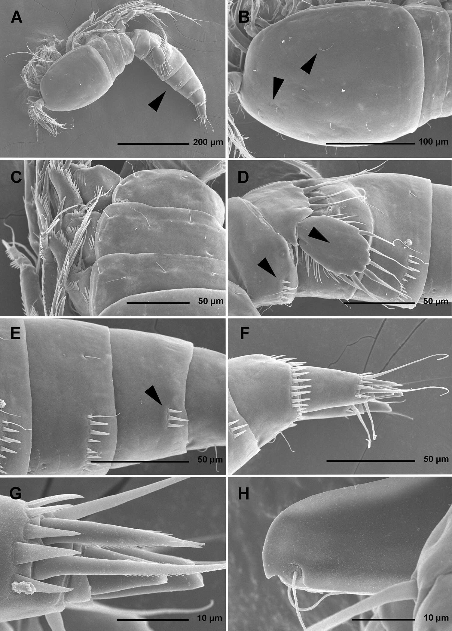
Figure 1.Stenhelia pubescens Chislenko, 1978, scanning electron micrographs, female 1: A habitus, lateral B cephalothorax, lateral C free thoracic somites, lateral D fifth pedigerous somite and genital double-somite, lateral, with one spermatophore attached on ventral side E fourth and fifth urosomites, lateral F anal somite and caudal rami, lateral G first legs and proximal part of second and third legs, lateral H distal part of right antennula, dorsal.
-
Tomislav Karanovic, Kichoon Kim, Wonchoel Lee
Zookeys
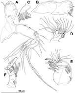
Figure 2.Stenhelia pubescens Chislenko, 1978, scanning electron micrographs, ovigerous female 2: A habitus, ventral B rostrum and left antennula, ventral C mouth appendages, ventral D first leg, anterior E second, third, and fourth legs, anterior F exopod of fifth leg and sixth leg, ventral G anal somite and caudal rami, ventral H posterior part of left caudal ramus, ventral.
-
Tomislav Karanovic, Kichoon Kim, Wonchoel Lee
Zookeys
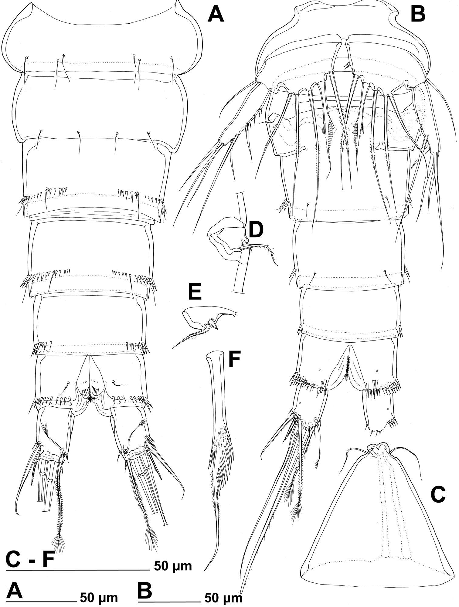
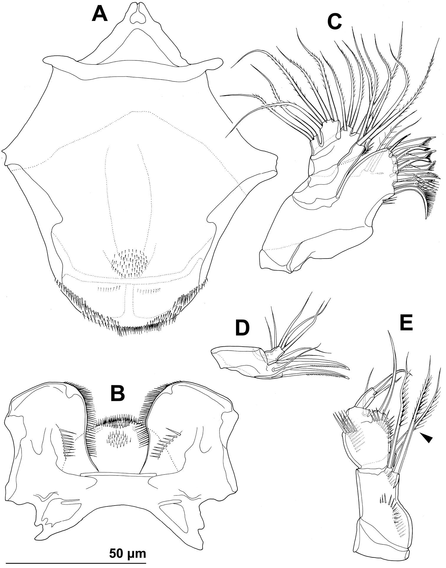
Figure 3.Stenhelia pubescens Chislenko, 1978, line drawings, female 3: A urosome, dorsal B urosome, ventral (armature on left caudal ramus omitted) C rostrum, dissected and compressed, dorsal D sixth leg, dorso-lateral E sixth leg, ventro-lateral F fifth leg second endopodal seta from inner side, anterior.
-
Tomislav Karanovic, Kichoon Kim, Wonchoel Lee
Zookeys
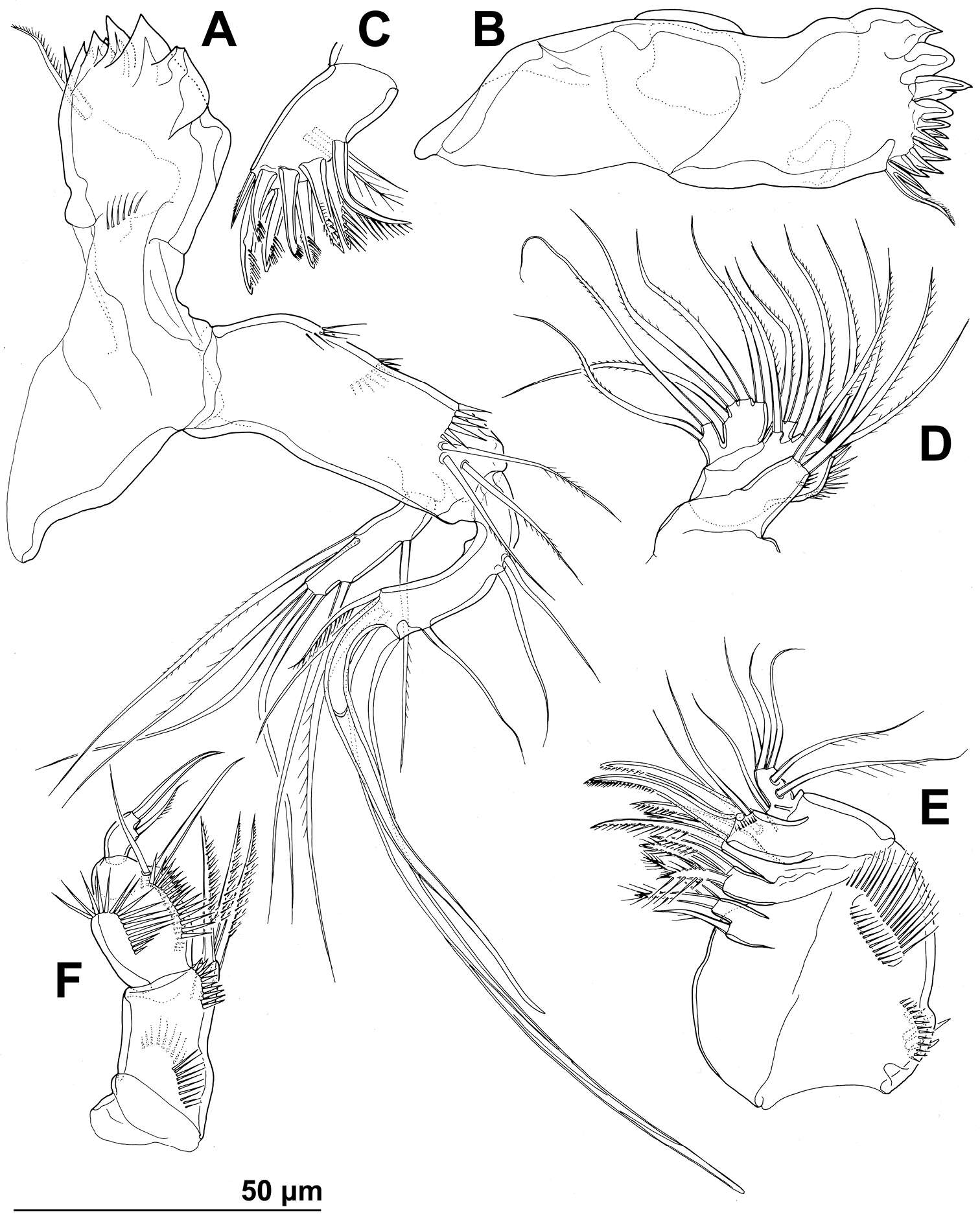
Figure 4.Stenhelia pubescens Chislenko, 1978, line drawings, female 3: A mandibula, posterior B mandibular coxa, anterior C maxillula, praecoxa arthrite, posterior D maxilular palp, posterior E maxilla, anterior F maxilliped, anterior.
-
Tomislav Karanovic, Kichoon Kim, Wonchoel Lee
Zookeys
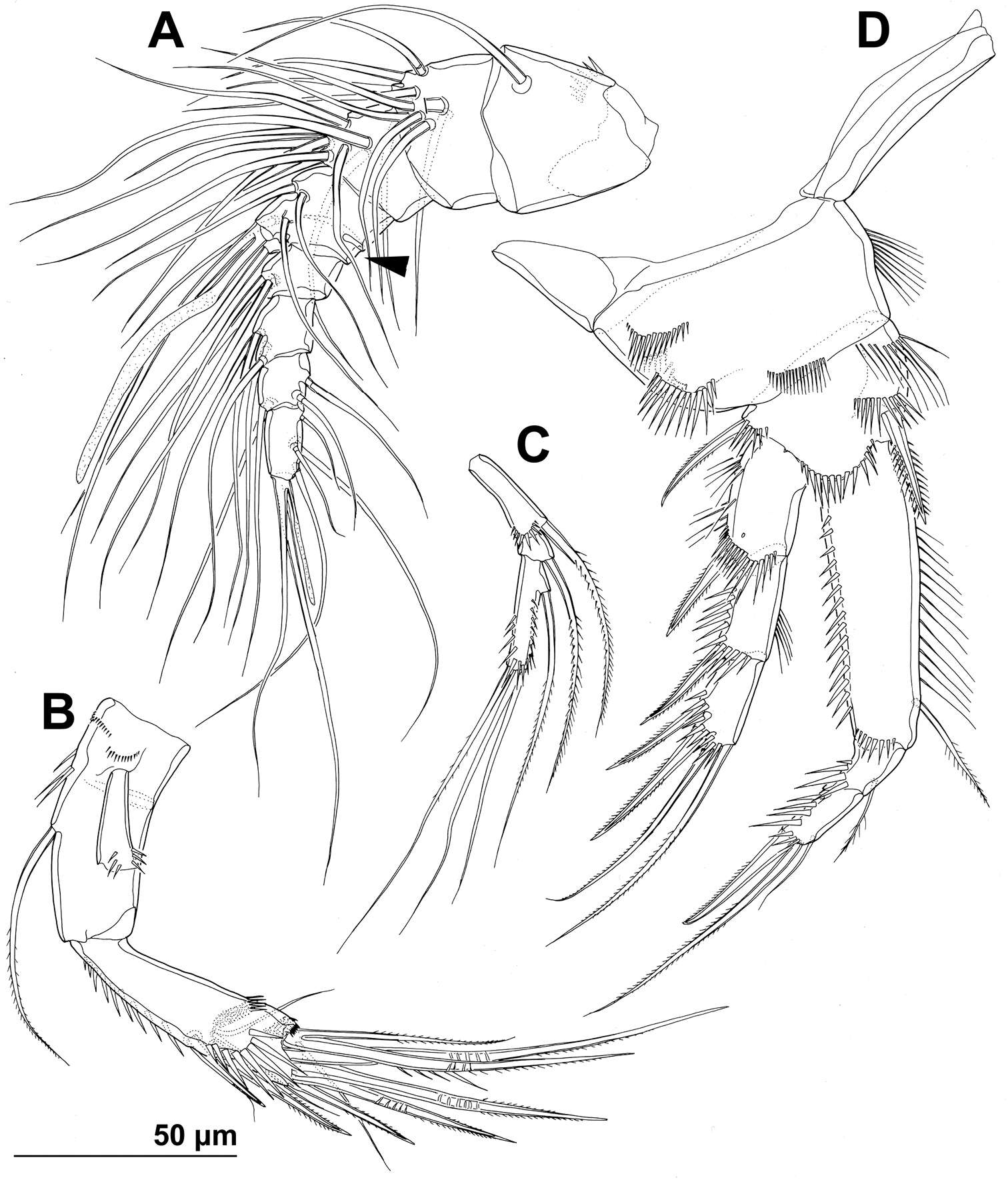
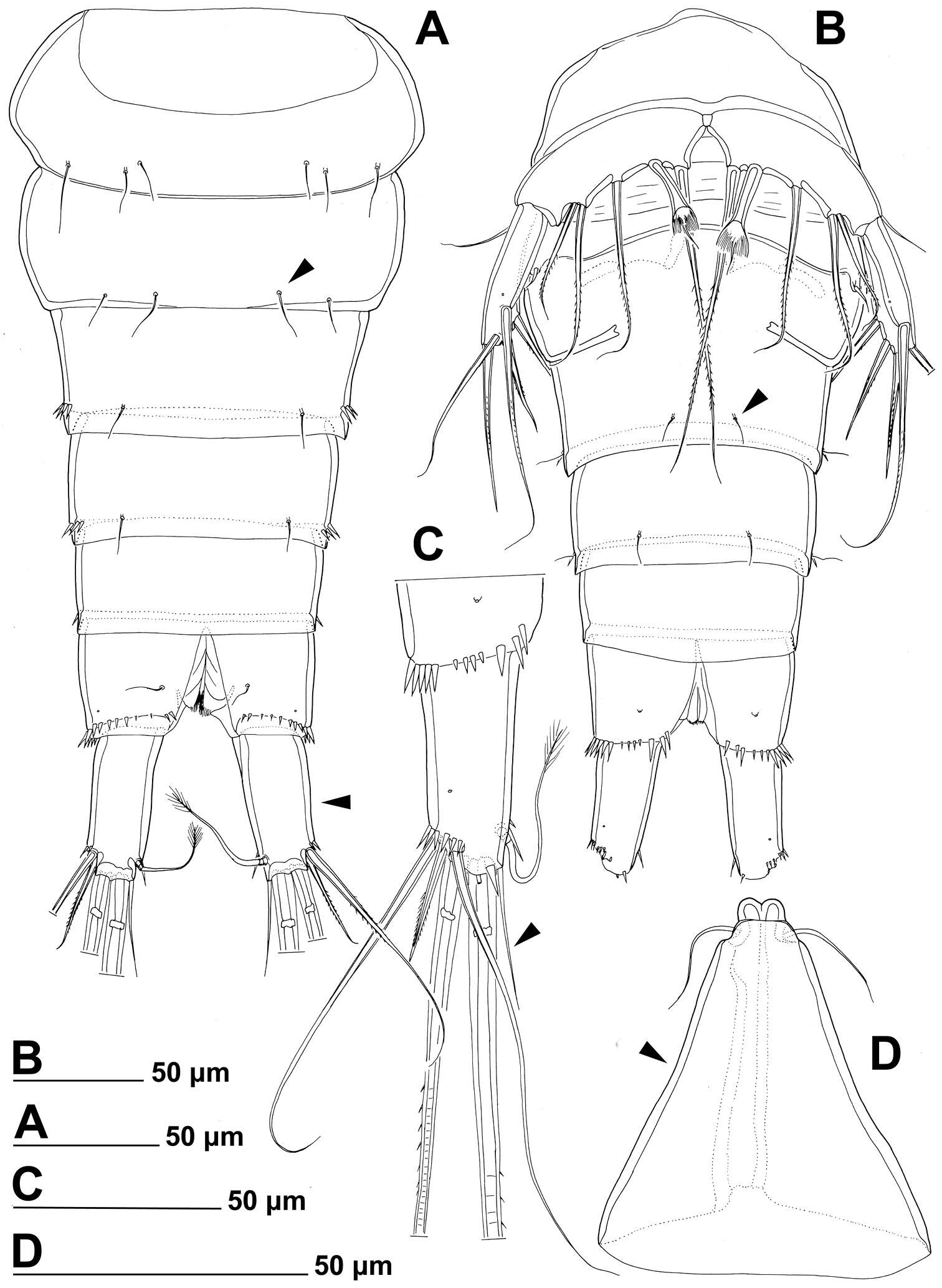
Figure 5.Stenhelia pubescens Chislenko, 1978, line drawings, female 3: A antennula, ventral B basis, endopod, and first exopodal segment of antenna, anterior C antennal exopod, anterior D first leg, anterior. Arrowhead indicates the presence of caudal suture on the fourth antennular segment.
-
Tomislav Karanovic, Kichoon Kim, Wonchoel Lee
Zookeys
Figure 6.Stenhelia pubescens Chislenko, 1978, line drawings, female 3: A second leg, anterior B third leg, anterior.
-
Tomislav Karanovic, Kichoon Kim, Wonchoel Lee
Zookeys
Figure 7.Stenhelia pubescens Chislenko, 1978, line drawings, female 3: A fourth leg, anterior B fifth leg, dissected and flattened, anterior.
-
Tomislav Karanovic, Kichoon Kim, Wonchoel Lee
Zookeys
Figure 7.Stenhelia pubescens Chislenko, 1978, line drawings, female 3: A fourth leg, anterior B fifth leg, dissected and flattened, anterior.
-
Tomislav Karanovic, Kichoon Kim, Wonchoel Lee
Zookeys
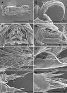
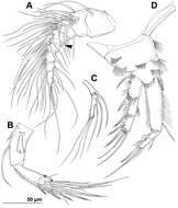
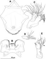
Figure 8.Stenhelia taiae Mu & Huys, 2002, scanning electron micrographs, female: A habitus, lateral B cephalothoracic shield, lateral C free thoracic somites, lateral D fifth pedigerous somite and genital double-somite, lateral E fourth and fifth urosomites, lateral F anal somite and caudal rami, lateral G posterior part of right caudal ramus, lateral H rostrum, lateral. Arrowheads indicate morphological characters different from those in Stenhelia pubescens Chislenko, 1978.
-
Tomislav Karanovic, Kichoon Kim, Wonchoel Lee
Zookeys
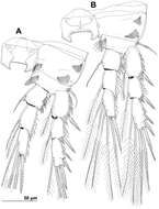
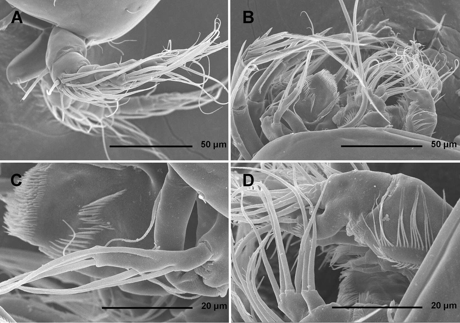
Figure 9.Stenhelia taiae Mu & Huys, 2002, scanning electron micrographs, female: A rostrum and antennulae, lateral B antenna and mouth appendages, lateral C mandibular palp and labrum, lateral D maxilla and part of maxillular palp, lateral.
-
Tomislav Karanovic, Kichoon Kim, Wonchoel Lee
Zookeys
Figure 10.Stenhelia taiae Mu & Huys, 2002, line drawings, female: A urosome, dorsal B urosome, ventral (caudal rami armature omitted) C right caudal ramus, ventral D rostrum, dissected and compressed, dorsal. Arrowheads indicate morphological characters different from those in Stenhelia pubescens Chislenko, 1978.
-
Tomislav Karanovic, Kichoon Kim, Wonchoel Lee
Zookeys
Figure 11.Stenhelia taiae Mu & Huys, 2002, line drawings, female: A labrum, posterior B paragnaths, anterior C maxillula, posterior D maxillar basis and endopod, posterior E maxilliped, posterior. Arrowhead indicates morphological character different from that in Stenhelia pubescens Chislenko, 1978.
-
Tomislav Karanovic, Kichoon Kim, Wonchoel Lee
Zookeys
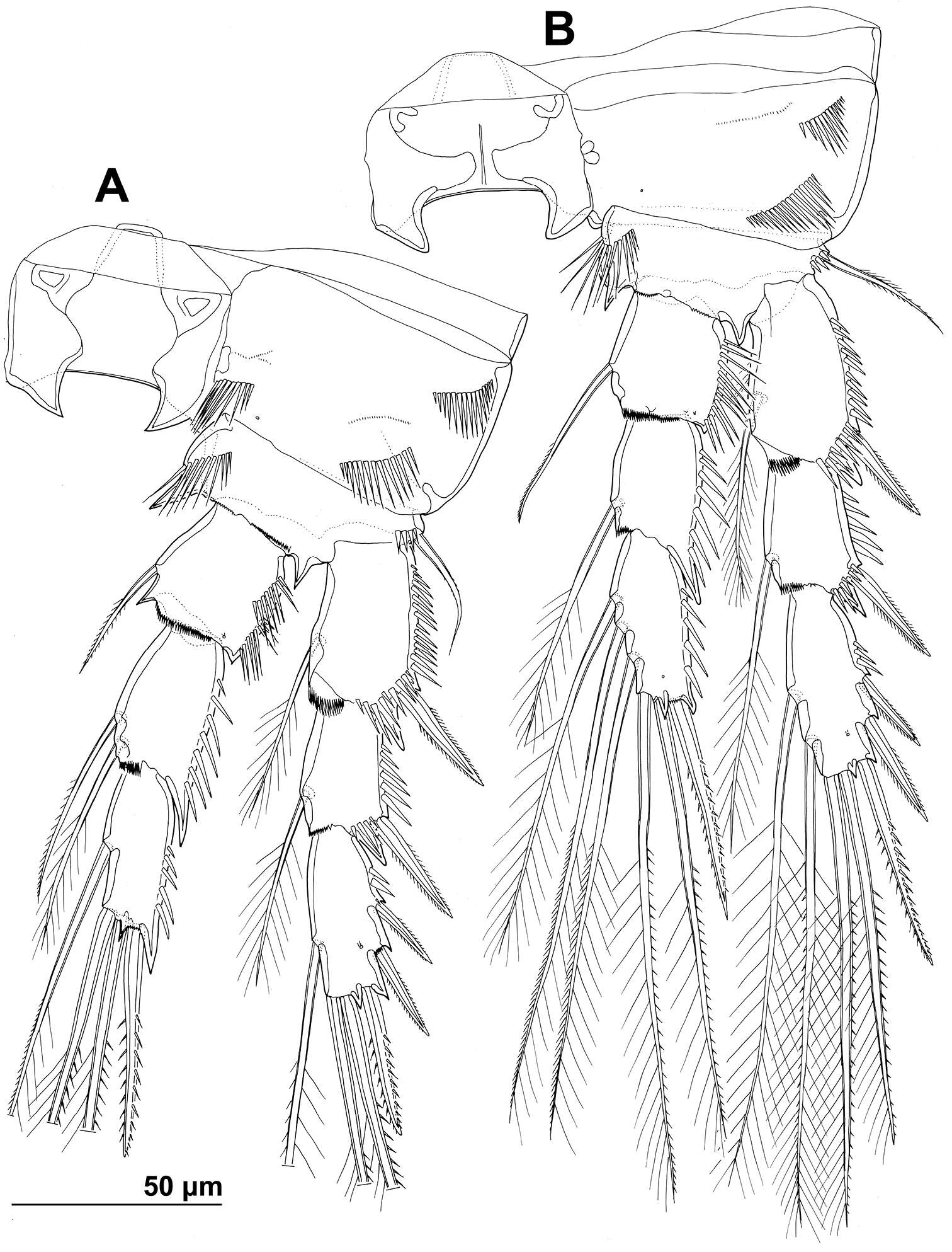
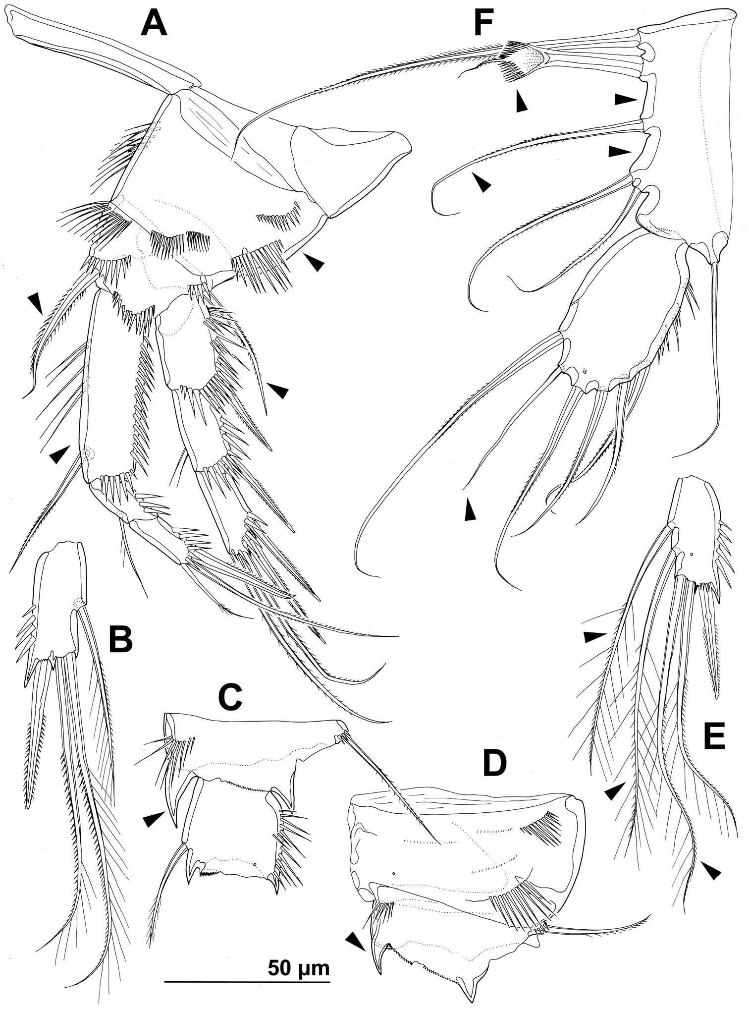
Figure 12.Stenhelia taiae Mu & Huys, 2002, line drawings, female: A first leg, anterior B third endopodal segment of second leg, anterior C basis and first endopodal segment of third leg, anterior D coxa and basis of fourth leg, anterior E third endopodal segment of fourth leg, anterior F fifth leg, dissected and flattened, anterior. Arrowheads indicate morphological characters different from those in Stenhelia pubescens Chislenko, 1978.
-
All Biocode files are based on field identifications to the best of the researcher’s ability at the time.
-
All Biocode files are based on field identifications to the best of the researcher’s ability at the time.
-
All Biocode files are based on field identifications to the best of the researcher’s ability at the time.
-
All Biocode files are based on field identifications to the best of the researcher’s ability at the time.
-
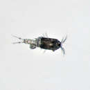
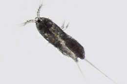
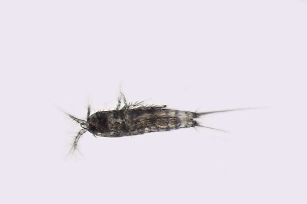
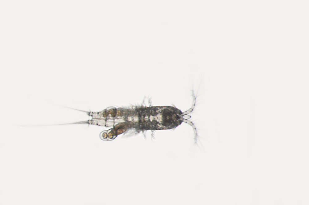
USNM 1499616 - Specimen Image; Field number: SERCINVERT2927
-
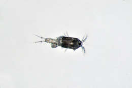
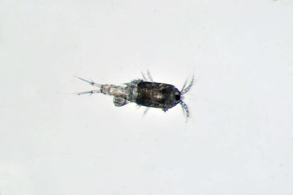
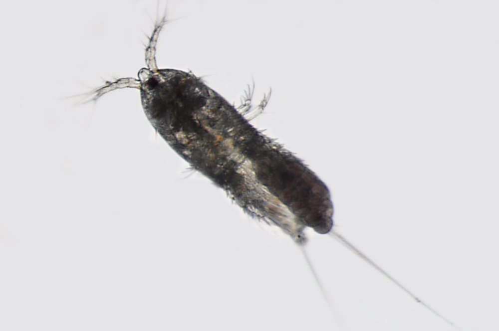
USNM 1499710 - Specimen Image; Field number: SERCINVERT3021