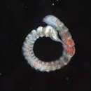
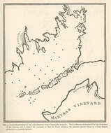
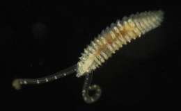
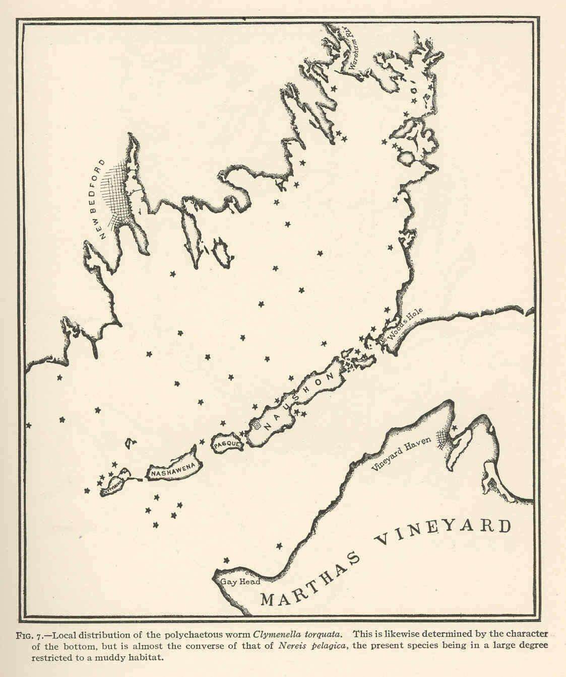
Local distribution of the polychaetous worm Clymenella torquata. This is likewise determined by the character of the bottom, but is almost the converse of that of Nereis pelagica, the present species being in a large degree restricted to a muddy habitat.
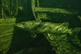
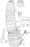
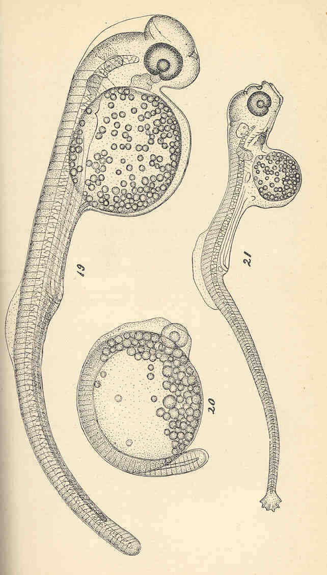
Siphostoma fuscum (The Pipe-Fish).. fig. 19Young embryo, in which the tail is still archicereal and the dorsal and pectoral fins are just developing. fig.20 a still younger stage, in which the tail is just beginning to grow out. fig.21 an older stage, in which the caudal fin is beginning to be formed
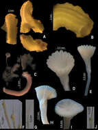
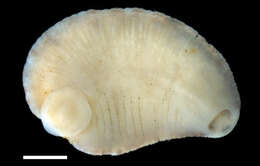
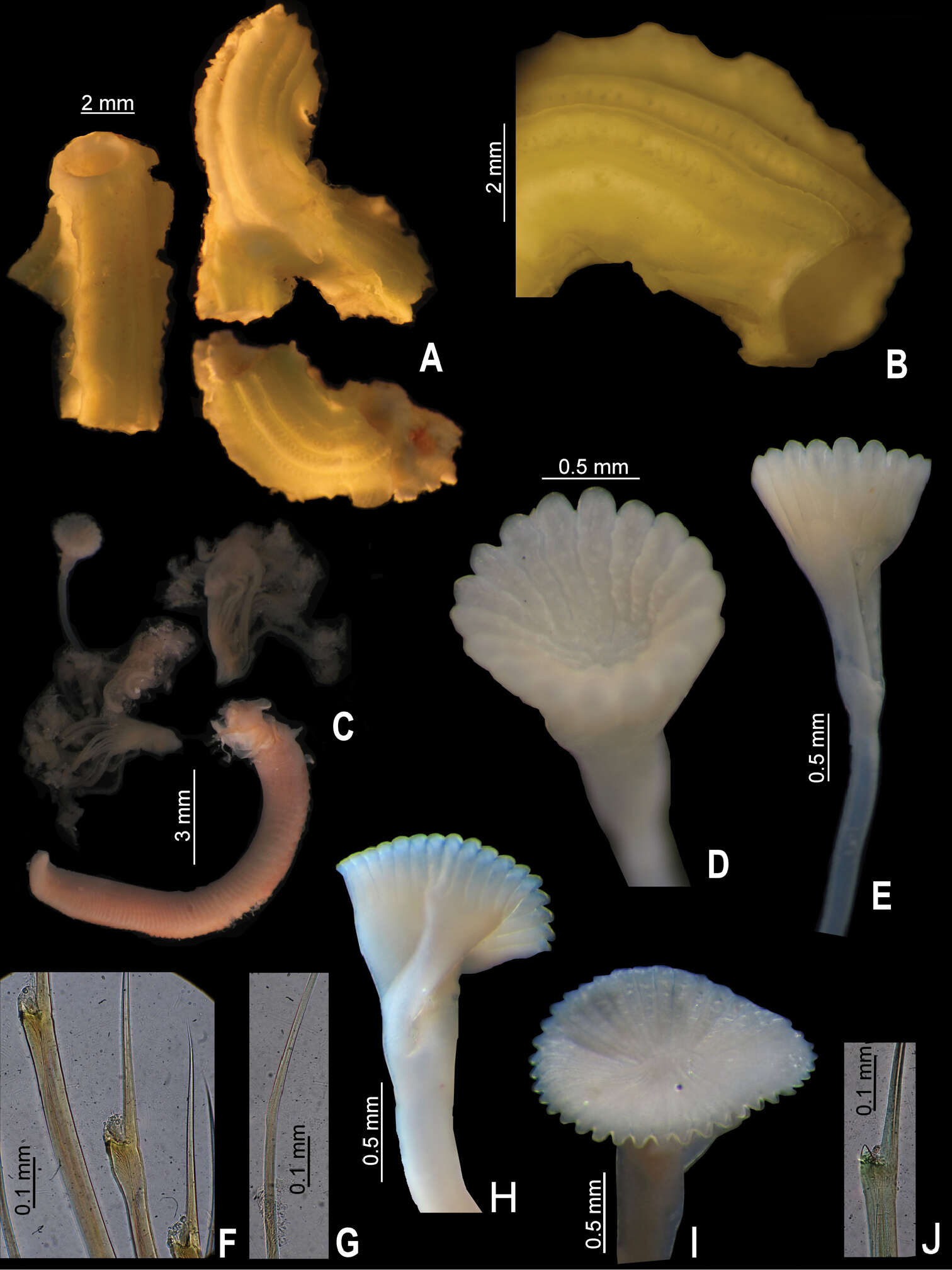
Figure 2.A–G Serpula madrigalae sp. n., from Turks and Caicos, USNM 1157006, holotype A–B tube and detail C entire body D–E operculum, in aboral and lateral views F bayonet chaetae G hooded (capillary) chaetae H–J Serpula cf. vermicularis, from Nigeria, UMML 22.545 H–I two distinct opercula in lateral and aboral views J bayonet chaetae.
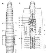
Figure 12.Morphology of the lectotype (ZMH V9293) of Glyphidrilus jacobsoni Michaelsen, 1922, showing the A external ventral and B internal dorsal views.
Figure 11.Sternaspis maior Chamberlin, 1919 A Neotype (UNAM 7882), ventral view B Same, lateral view C Same, anterior end, frontal view D Same, ventro-caudal shield E Paraneotype (UNAM Sta. 13), ventro-caudal shield F Paraneotype (UNAM Sta. 13, OH), ventro-caudal shield G Paraneotype (UNAM 7881), ventro-caudal shield. Sternaspis princeps Selenka, 1885, syntypes (NHM 1885.12.3.1) H Larger syntype, median region showing gonopodial lobes I Smaller syntype, ventro-caudal shield, frontal view. Bars: A 1.9 mm B 2 mm C 1 mm D 1.4 mm E 1.3 mm F 1.5 mm G 2.5 mm H, I 1.2 mm.
Robert J. Blakemore, Seunghan Lee, Wonchoel Lee, Hong-Yul Seo
Zookeys
Figure 2.Amynthas jinburi sp. n. showing ventral view with spermathecae and 18rhs prostate in situ plus simple intestinal caecum in 27; dorsal view of prostomium; [boxed is 2X lateral view of spermathecal pores in 5 & 6rhs].
Sergio I. Salazar-Vallejo, Galina Buzhinskaja
Zookeys
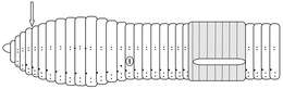
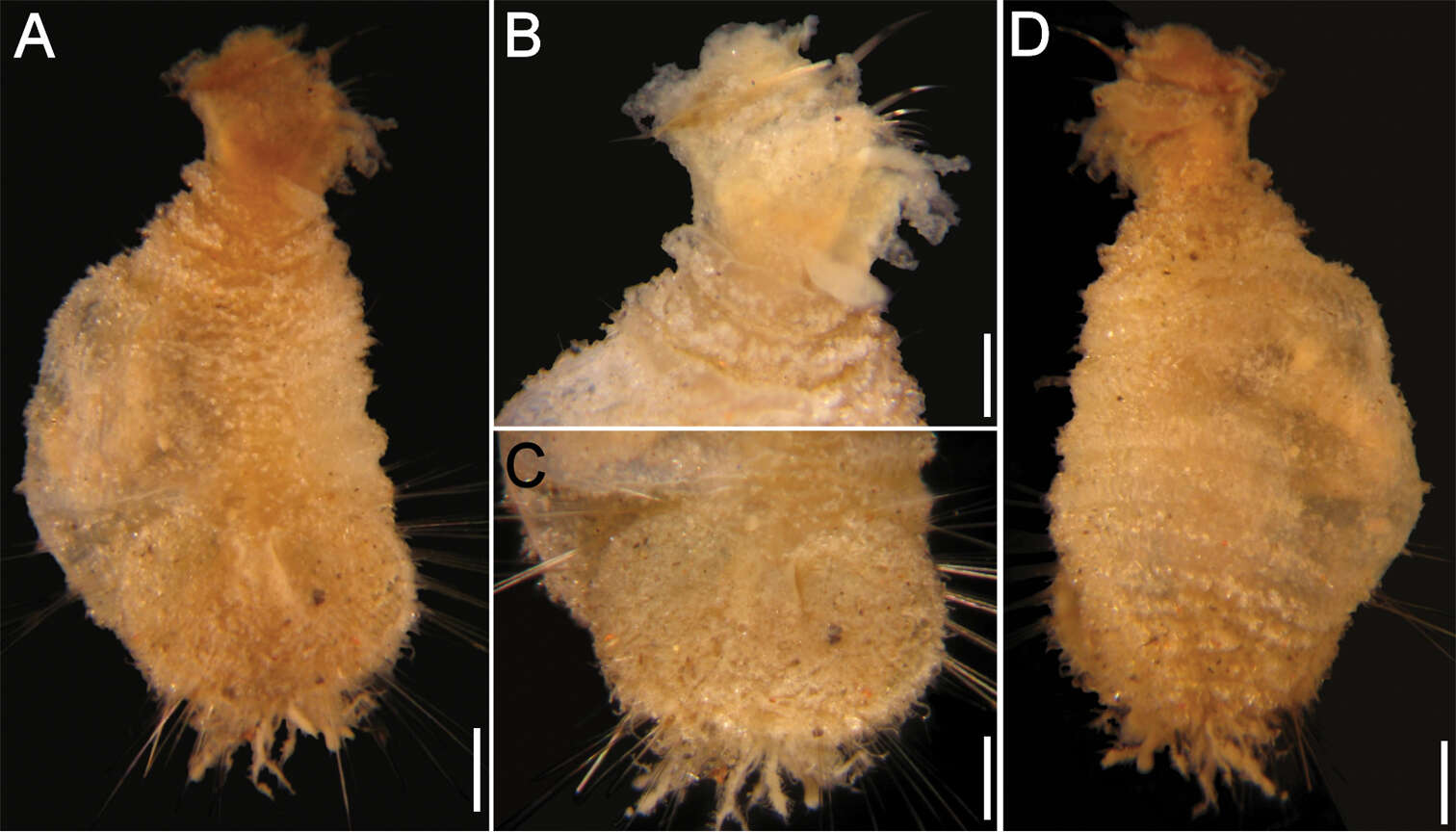
Figure 2.Caulleryaspis fauchaldi sp. n., juvenile specimen (LACM 5362) A Ventral view B Anterior end, ventral view C Ventro-caudal shield, frontal view D Dorsal view. Bars: A, C, D 0.38 mm B 0.26 mm.
María E. García-Garza, J.A. de León-González
Zookeys
Figure 3.Amphictene guatemalensis. Holotype. A ventral view of anterior end B dorsal view of scaphe D scaphal hooks. Bar scale = A, B = 1 mm; C = 20mm.
William A. Hopkins, William E. Moser, David W. Garst, Dennis J. Richardson, Charlotte I. Hammond, Eric A. Lazo-Wasem
Zookeys
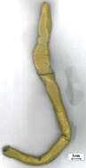
Figure 3.Ventral surface of Placobdella appalachiensis sp. n., Holotype USNM 1232924 collected from an adult eastern hellbender (Cryptobranchus alleganiensis) from stream reach A3 in southwest Virginia, USA. Scale bar equals 1 mm.
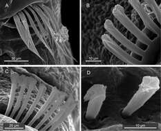
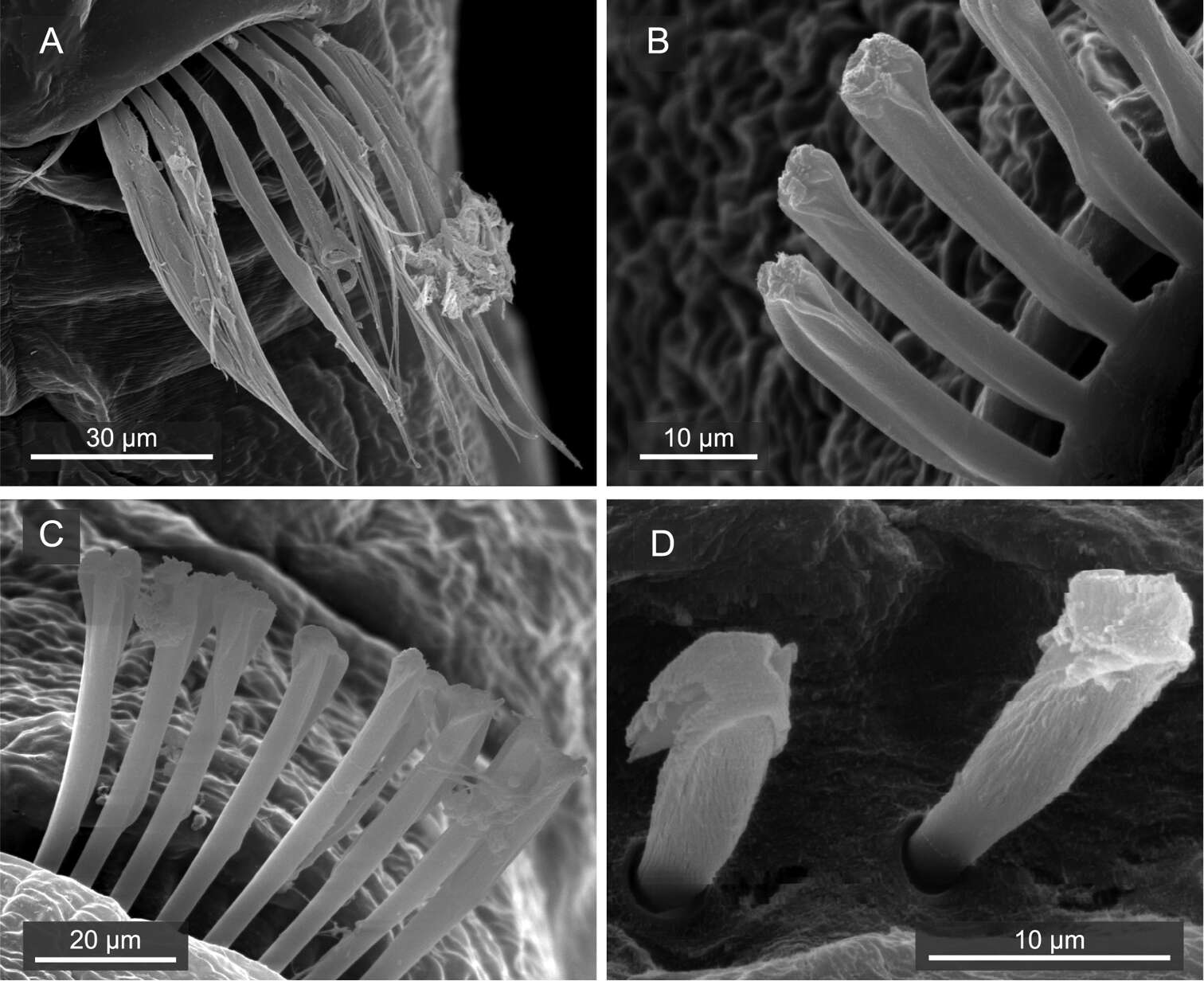
Figure 3.Mediomastus duobalteus sp. n., paratype, CBM-ZW 1089, SEM images. A Capillary chaetae on segment 3 B notopodial hooded hooks on segment 6 C neuropodial hooded hooks on segment 6 D notopodial hooded hooks on segment 13.
Ueangfa Bantaowong, Ratmanee Chanabun, Piyoros Tongkerd, Chirasak Sutcharit, Samuel W. James, Somsak Panha
Zookeys
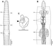
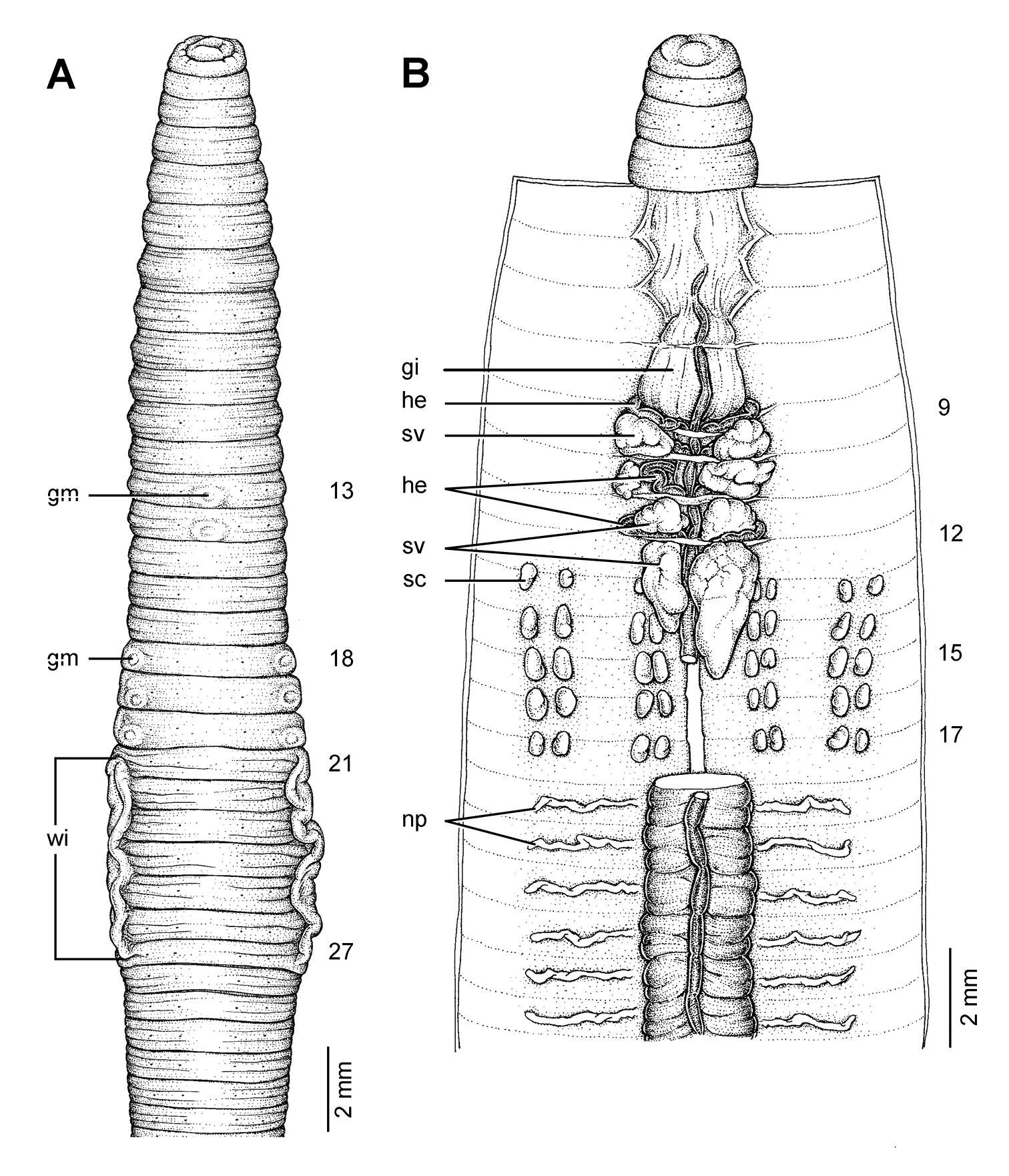
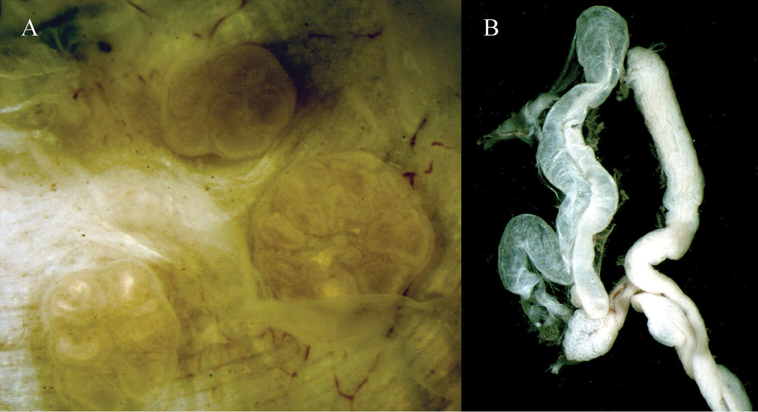
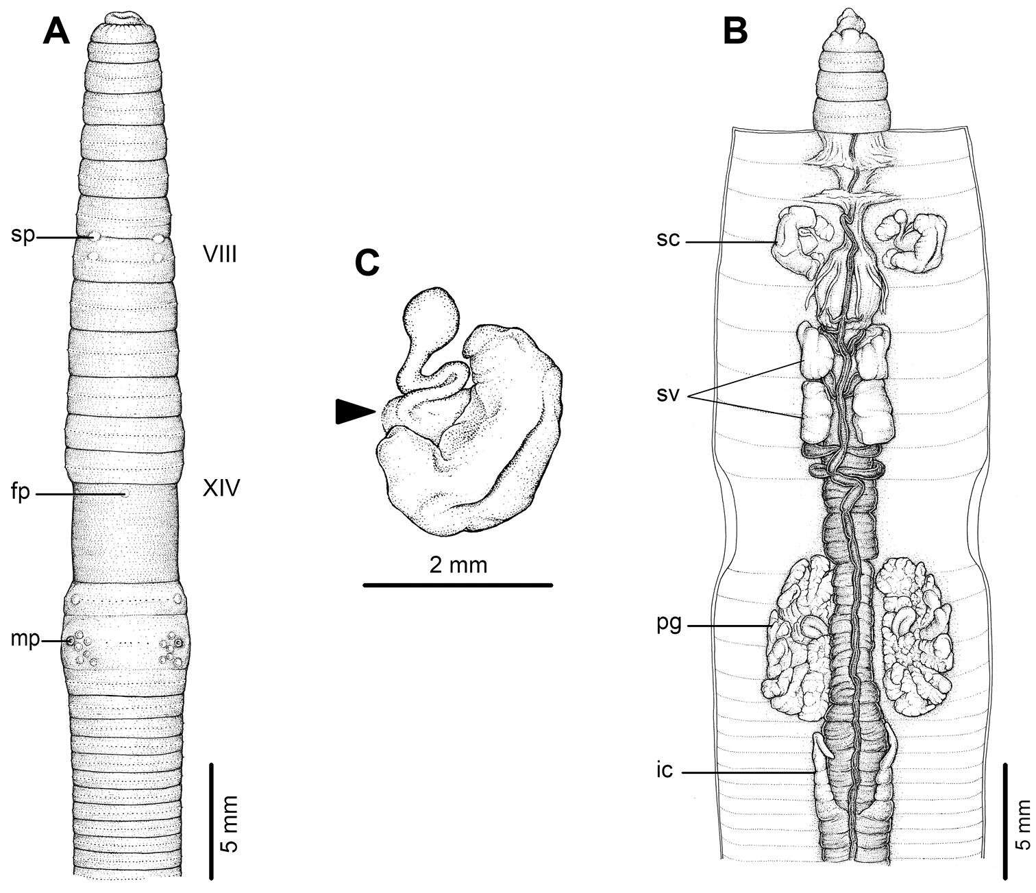
Figure 2.External and internal morphology of holotype (CUMZ 3204) of Amynthas phatubensis sp. n. A External ventral view, B internal dorsal view and C spermatheca, and black arrow indicates the connection of the spermatheca and spermathecal pore.