-
-
-
-
-
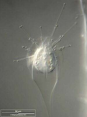
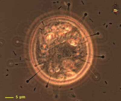
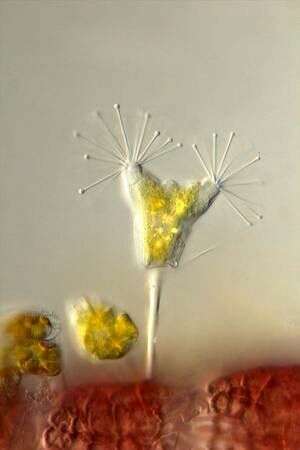
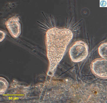
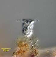
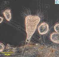
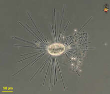
Portrait of Metacineta BÃTSCHLI,1889. Suctorian residing in vase shaped lorica with slit-like apertures through which feeding tentacles protrude. Knob-like swellings of haptocysts are seen at the ends of the tentacles. These serve to immobilize and hold prey whose contents are transported down a microtubular channel in the tentacle to a food vacuole in the cell body. From freshwater pond near Boise, Idaho. Phase contrast
-
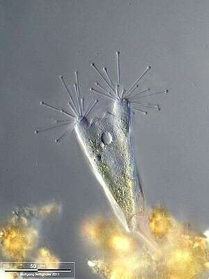
Portrait of Metacineta BÃTSCHLI,1889. Suctorian residing in vase shaped lorica with slit-like apertures through which feeding tentacles protrude. Knob-like swellings of haptocysts are seen at the ends of the tentacles. These serve to immobilize and hold prey whose contents are transported down a microtubular channel in the tentacle to a food vacuole in the cell body. From freshwater pond near Boise, Idaho. DIC
-
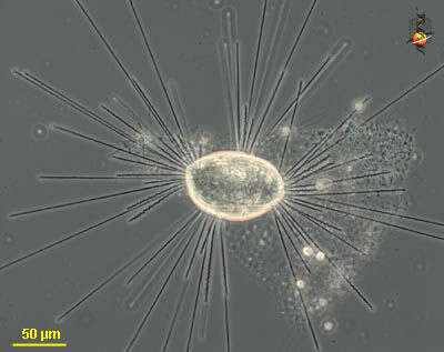
Scale bar indicates 50 µm. Sample from a pond situated in the vicinity of Lake Constance (Bodensee, Southern Germany). The image was built up using several photomicrographic frames with manual stacking technique. Images were taken using Zeiss Universal with Olympus C7070 CCD camera.
-
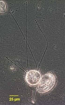
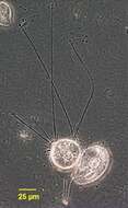
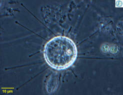
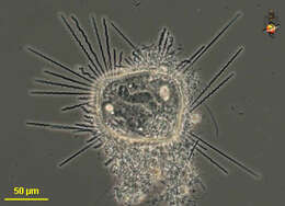
Prodiscophrya (pro-disc-owe-fry-a) is a suctorian (ciliate without cilia in the trophic stage but with multiple elongated mouths = arms). Arms expanded at apex where they are filled with extrusomes which are used in food capture. Arms not arranged in groups, usually stalked but this cell was observed without a stalk. Phase contrast.
-
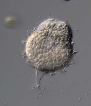
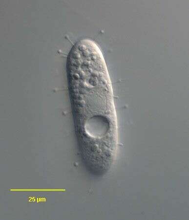
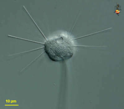
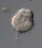
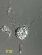
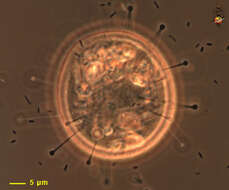
This image shows a recently settled cell. There are tentacles which are swollen at the apices. The surface of the cell has a few cilia - remaining from the swimming larva that settled to produce this feeding form. Phase contrast microscopy.
-
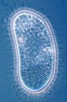
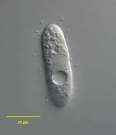
Ciliated larval stage of the suctor. Phase contrast micrograph of living cell.
-
-
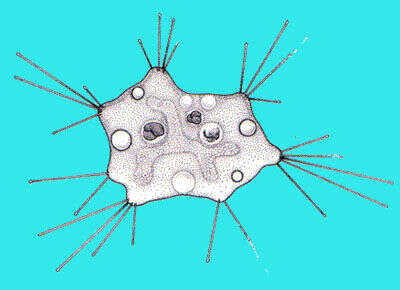
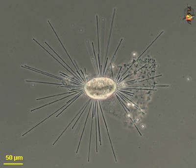
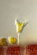
Solenophrya, suctorian that occurs adpressed to the substrate, arms extend in all direction. Each arm is a mouth, the ip of which is expanded with the extrusomes which help it capture its food. From Lake Donghu, China. Phase contrast micrograph.
-
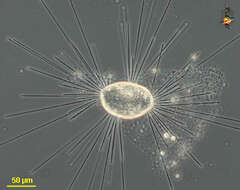
in vivo portrait of the suctorian, Acineta tuberosa (Ehrenberg, 1834). The capitate tentacles protrude from two conical protruberances called actinophores. A contractile vacuole is visible. Collected from a commercial saltwater aquarium in Boise, Idaho. DIC.
-
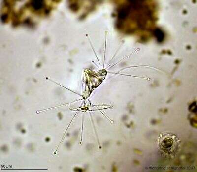
This picture made with water immersion objective without using a cover glass provides an apical look on an undisturbed Acineta. Collected from "Bodden", the brackish waters lying between the isles of Hiddensee and Ruegen (German Baltic Sea). This multi layer image (DOF)was built up using 10 DIC frames with manual stacking technique using Corel Photopaint. The scale bar indicates 50 µm. Images were taken using Zeiss Universal with Olympus C7070 CCD camera.
-
Scale bar indicates 50 µm. Sample from a pond situated in the vicinity of Lake Constance (Bodensee, Southern Germany). The image was built up using several photomicrographic frames with manual stacking technique. Images were taken using Zeiss Universal with Olympus C7070 CCD camera.
-
Collected from Bodden, the brackish waters lying between the isles of Hiddensee and Ruegen (German Baltic Sea).Images were taken using Zeiss Universal with Canon 600D.Image under Creative Commons License V 3.0 (CC BY-NC-SA).
-
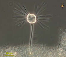
Tokophrya (toke-owe-fry-a) is a suctorian ciliate (there are no cilia in the trophic form, but multiple elongate mouths usually referred to as arms). The arms are arranged in groups (here a cluster to each side, but none across the middle). Attached to the substrate by a stalk. Phase contrast.
-
Tokophrya (toke-owe-fry-a) is a suctorian ciliate (there are no cilia in the trophic form, but multiple elongate mouths usually referred to as arms). The arms are arranged in groups (here a cluster to each side, but none across the middle). Attached to the substrate by a stalk. Phase contrast.
-
-
Tokophrya, a stalked suctorian ciliate. Cells with a stalk but no lorica. Arms form groups (fascicles) at the anterior end. Commonly found among peritrich colonies in Lake Donghu, China. Phase contrast micrograph.
-
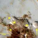
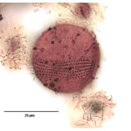
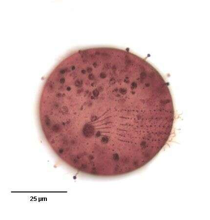
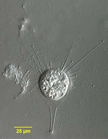
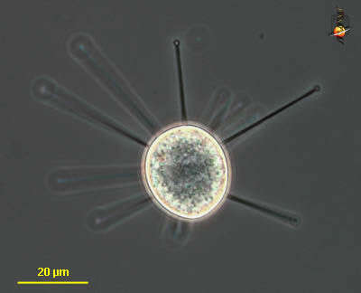
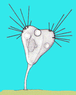
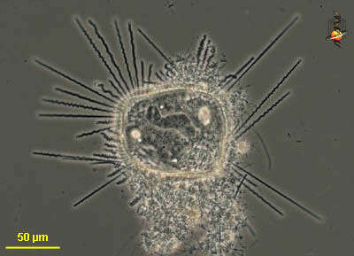
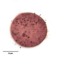
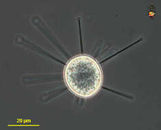
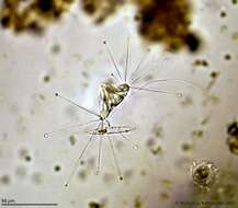
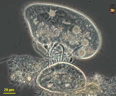
Heliophrya (heal-ee-owe-fry-a) is a stalkless suctorian, the body is a flattened disc to square shape, adpressed to the substrate, with contractile arms emerging in a number (usually 4) bundles. Food, mostly ciliates, is caught on the ends of the arms. The tips of the arms are swollen with extrusomes etc. which are used in food capture. Phase contrast.
-
Heliophrya (heal-ee-owe-fry-a) is a stalkless suctorian, the body is a flattened disc to square shape, adpressed to the substrate, with contractile arms emerging in a number (usually 4) bundles. This image shows the contracting arms with the central dark component being the microtubules of the mouth (each arm is a mouth). Phase contrast.
-
Heliophrya (heal-ee-owe-fry-a) is a stalkless suctorian, the body is a flattened disc to square shape, adpressed to the substrate, with contractile arms emerging in a number (usually 4) bundles. Food, mostly ciliates, is caught on the ends of the arms. The tips of the arms are swollen with extrusomes etc. which are used in food capture. Phase contrast.
-
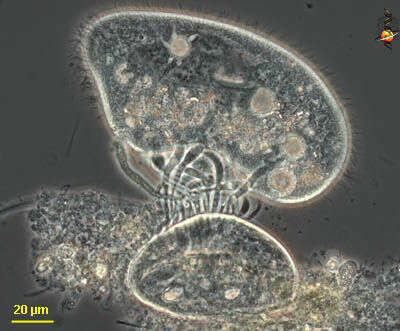
Heliophrya (heal-ee-owe-fry-a) is a stalkless suctorian, the body is a flattened disc to square shape, adpressed to the substrate, with contractile arms emerging in a number (usually 4) bundles. As can be seen here, food is caught on the ends of the arms, the arms act as mouths and the cytoplasm of the living prey (the prey is Paramecium and the contractile vacuoles are still active) is sucked down the arms and into the suctorian. Phase contrast.