-
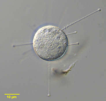
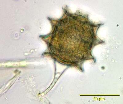
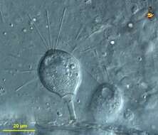
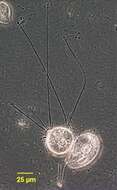
Suctoria are predatory ciliates. However, they give very little indication of the ciliate-ness. There are normally no cilia. Suctoria are sessile, and only when they divide do they produce ciliated forms which then swim away and find somewhere new to settle. Instead the body produces a number of tentacles. These are mouths, and the end of the them are expanded because of a concentration of extrusomes which are used to grab hold of their food - usually other protists. This form is stalked. Differential interference contrast.
-
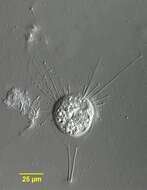
Suctoria are predatory ciliates. However, they give very little indication of the ciliate-ness. There are, for example, normally no cilia. Instead the body produced a number of tentacles. These are mouths, and the end of the them are expanded because of a concentration of extrusomes which are used to grab hold of their food - usually other protists. Suctoria are sessile, and only when they divide do they produce ciliated forms which then swim away and find somewhere new to settle. This form is stalked. Phase contrast.
-
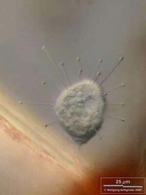
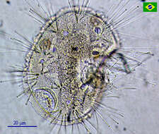
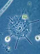
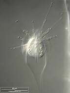
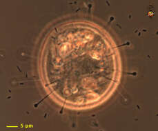
An unidentified suctorian. Very interesting ciliates, the Suctoria are provided with tentacles and rarely shows up in samples from the polluted Tiete River.
-
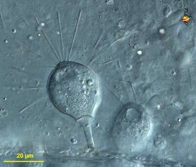
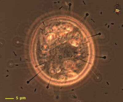
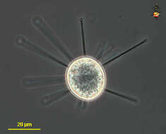
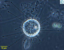
Portrait of Sphaerophrya, a spherical suctorian, which floats, free without lorica or stalk. There are evenly distributed capitate tentacles over the body surface. The tentacles retract with a typical accordion appearance (seen in this image at the 2 o clock position). There is a large centrally placed coarsely granular macronucleus and a single contractile vacuole. Some species are parasitic. Sphaerophrya preys on ciliates. From standing freshwater in Typha (cattail) marsh near Boise, Idaho. Differential interference contrast. Differential interference contrast optics.
-
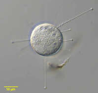
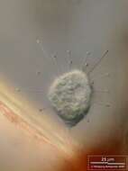
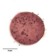
Small disc-shaped suctorian, usually adpressed to the substrate. With radiating arms. Phase contrast image of living cell.
-
-
Suctor Podophrya spec. living epibiotic on the red alga Ceramium. Collected from Bodden, the brackish waters lying between the isles of Hiddensee and Ruegen (German Baltic Sea). This image was taken using Zeiss Universal with Olympus C7070 CCD camera.
-
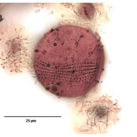
Typical biocoenosis of Suctoria (Podophrya spec.) with Peritricha on filaments of the red alga Ceramium diaphanum . Peritricha (not only the ones with lorica) don´t act as prey for the Suctoria. Scale bar indicates 25 µm. Collected from Bodden, the brackish waters lying between the isles of Hiddensee and Ruegen (German Baltic Sea). This image was taken using Zeiss Universal with Olympus C7070 CCD camera.
-
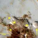
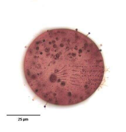
Podophrya spec. hast trapped a Trichodina and is ingesting their cytoplasm. There ist no suction as the name "Suctoria" indicates misleadingly. An active membrane transport based on a microtubular system inside the tentacles is the engine of ingestion of the prey´s cytoplasm. The ingesting tentacles are widened. Collected from Bodden, the brackish waters lying between the isles of Hiddensee and Ruegen (German Baltic Sea). This image was taken using Zeiss Universal with Olympus C7070 CCD camera.
-
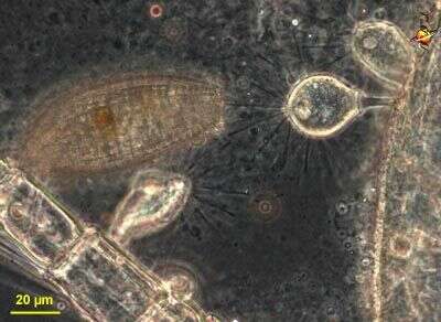
Portrait of ciliated swarmer or larval form of the suctorian, Podophrya fixa (MUELLER, 1786) EHRENBERG, 1833. Cilia are seen interspersed with retracted capitate tentacles. The adult form is very similar in appearance to Prodiscophrya but swarmer form of Podophrya has only one contractile vacuole while that of Prodiscophrya has two. The swarmer secretes a long rigid hollow stalk, which attaches the adult to the substrate by an adhesive disc. In the adult form tentacles remain while cilia disappear. The spheroid macronucleus is seen here. . The ciliated larval or swarmer form develops by budding. Podophrya may form a unique transversely ringed stalked resting cyst. Found in sapropelic habitats. From organically enriched bottom sediment of freshwater pond near Boise, Idaho. DIC optics.
-
Portrait of the stalked resting cyst of Podophrya fixa (MUELLER,1786) EHRENBERG, 1833, a suctorian ciliate. The thick brownish cyst wall has a variable (3-9) number of raised transverse rings. There is an apical aperture. The cell body is visible through the translucent cyst wall.
-
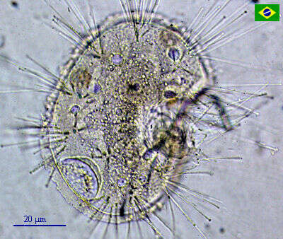
Portrait of adult form of the suctorian, Podophrya fixa (MUELLER,1786) Ehrenberg,1833. The cell body of the adult form has a spherical cell body atop a slender, hollow, rigid stalk that attaches to the substrate by an adhesive disc (seen here). There are numerous retractile capitate tentacles distributed over the entire cell surface. The knobs at the ends of the tentacles are aggregates of specialized extrusomes called haptocysts. These fix prey (usually ciliates) the contents of which are then transported to the cell body through the tentacles. No lorica. The adult form is very similar in appearance to Prodiscophrya but swarmer form of Podophrya has only one contractile vacuole while that of Prodiscophrya has two. The granular, spheroid macronucleus is central. The ciliated larval or swarmer form develops by budding. Podophrya may form a unique transversely ringed stalked resting cyst. Found in sapropelic habitats. From organically enriched bottom sediment of freshwater pond near Boise, Idaho. DIC optics.
-
-
-
-
-
Portrait of Metacineta BÃTSCHLI,1889. Suctorian residing in vase shaped lorica with slit-like apertures through which feeding tentacles protrude. Knob-like swellings of haptocysts are seen at the ends of the tentacles. These serve to immobilize and hold prey whose contents are transported down a microtubular channel in the tentacle to a food vacuole in the cell body. From freshwater pond near Boise, Idaho. Phase contrast
-
Portrait of Metacineta BÃTSCHLI,1889. Suctorian residing in vase shaped lorica with slit-like apertures through which feeding tentacles protrude. Knob-like swellings of haptocysts are seen at the ends of the tentacles. These serve to immobilize and hold prey whose contents are transported down a microtubular channel in the tentacle to a food vacuole in the cell body. From freshwater pond near Boise, Idaho. DIC
-
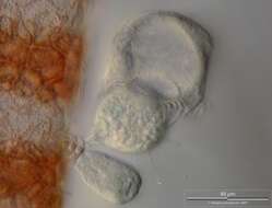
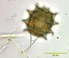
Scale bar indicates 50 µm. Sample from a pond situated in the vicinity of Lake Constance (Bodensee, Southern Germany). The image was built up using several photomicrographic frames with manual stacking technique. Images were taken using Zeiss Universal with Olympus C7070 CCD camera.
-
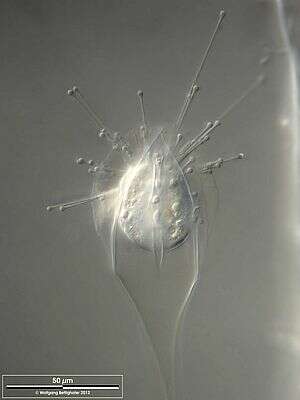
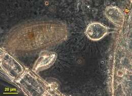
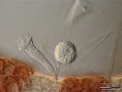
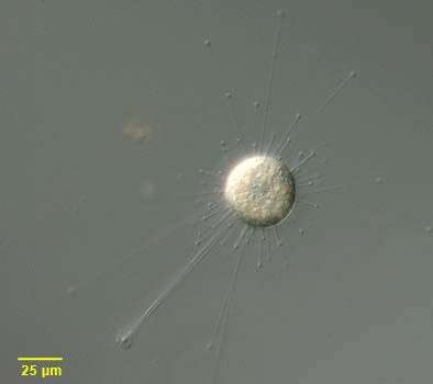
Prodiscophrya (pro-disc-owe-fry-a) is a suctorian (ciliate without cilia in the trophic stage but with multiple elongated mouths = arms). Arms expanded at apex where they are filled with extrusomes which are used in food capture. Arms not arranged in groups, usually stalked but this cell was observed without a stalk. Phase contrast.
-
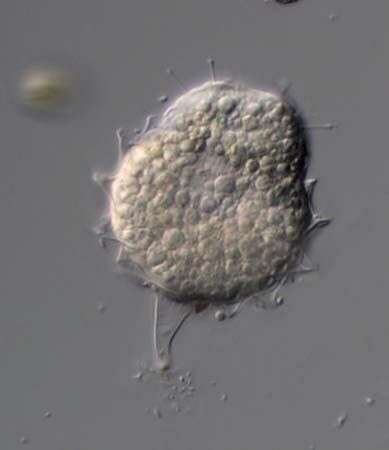
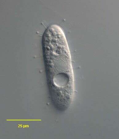
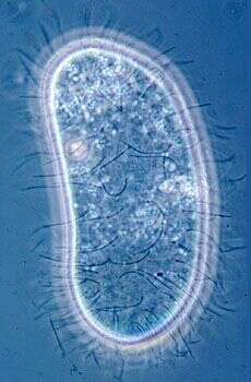
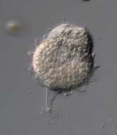
This image shows a recently settled cell. There are tentacles which are swollen at the apices. The surface of the cell has a few cilia - remaining from the swimming larva that settled to produce this feeding form. Phase contrast microscopy.
-
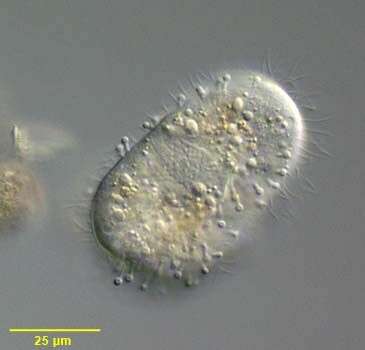
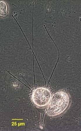
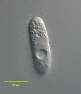
Ciliated larval stage of the suctor. Phase contrast micrograph of living cell.
-
-
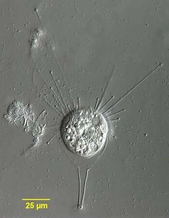
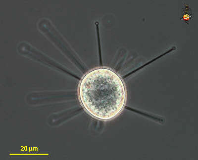
Solenophrya, suctorian that occurs adpressed to the substrate, arms extend in all direction. Each arm is a mouth, the ip of which is expanded with the extrusomes which help it capture its food. From Lake Donghu, China. Phase contrast micrograph.