-
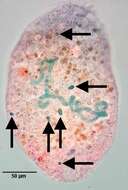
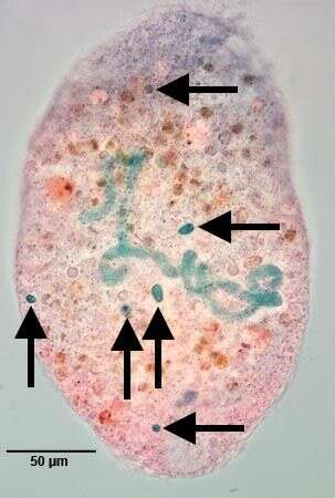
Six of the multiple (7-18) micronuclei are in the focal plane of this image. Stained by the methylgreen-pyroninY technique (see Foissner, W.Europ. J. Protistol.27:313-330;1991).Brightfield.
-
Pera, Faro, Portugal
-
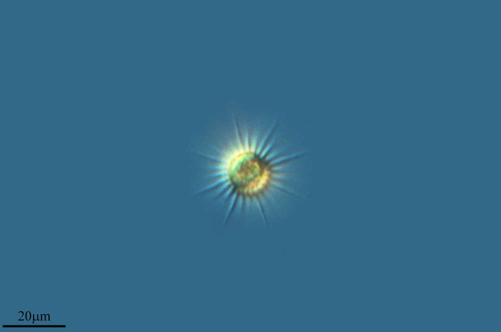
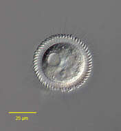
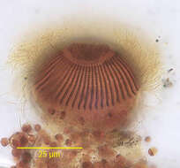
Pelagovasicola (pee-ladge-o-vee-sick-o-la) cinctum is a very fast swimming obovoid ciliate measuring 50 - 180 X 40 - 85 microns. It is common in plankton of lakes and ponds. The body is surrounded by 5-7 distinct ciliary girdles. The posterior fifth of the cell is unciliated. The contractile vacuole lies in the posterior end and has about 20 radial collecting channels. The macronucleus is kidney-shaped and lies in the mid-body. Extrusomes are arranged in the margin of the oral dome, occasionally extruded as bundles of fine filaments. This slightly squashed specimen was collected in the plankton of a bog pond near Konstanz, Germany, and this images emphasizes the radial collecting channels of the contractile vacuole. Differential interference contrast.
-
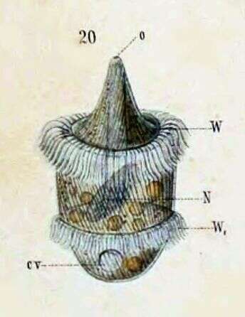
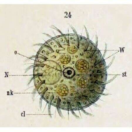
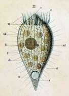
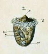
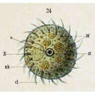
Originally described by Schewiakoff under the name Didinium balbianii. Schewiakoff describes this as an individual apprehended in division. o--Mouth N--Macronucleus W--Ciliated ring W1--New ciliated ring, prepared for offspring
-
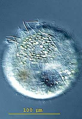
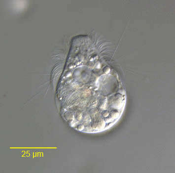
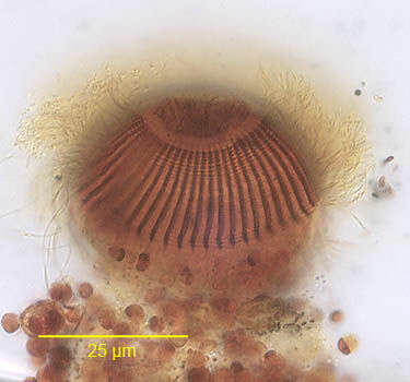
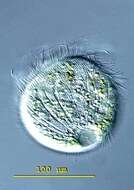
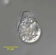
Posterior apical view of the haptorid ciliate,Askenasia volvox (Eichwald,1852) Kahl, 1930. The cell is spherical posteriorly and the anterior is a truncate cone.The cytostome is at the anterior apex.The cytostome is surrounded by an undulating line of granules (seen only in silver impregnated specimens).Somatic cilia are arranged in three (anterior,middle and posterior)girdles.The posterior girdle consists of long stiff bristles (seen here).The anterior cilia are directed forward and the middle girdle cilia are longer,curving backwards in a "sickle" configuration.These cilia produce the saltatory locomotion typical of this genus (they are seen well in this image).The posterior of the cell is unciliated.The central macronucleus is C-shaped. There is a single subequatorial contractile vacuole (seen here to viewer's left). From a freshwater pond near Boise, Idaho.DIC.
-
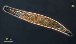
Homalozoon, a elongate-ribbon like predatory ciliate. The body is truncated (cut) off at the front end where the mouth is located, and pointed posteriorly. It has rows of cilia mostly on the ventral side, it glides over the substrate, sometimes contracting. Feeds on detritus and other protists. This image shows the macronucleus, which takes to form of a row of ellip[tical beads attached end-to-end. The micronuclei are small round dark structures adjacent to the macronucleus. Phase contrast micrograph.
-
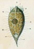
Originally described as Dinophrya liberkuhnii (Butschli) a -- Anus cl -- Cilia cv -- Contractile vacuole ek -- Ectoplasm N -- Macronucleus ncl -- Micronucleus nk -- Food particle o -- Mouth nk -- Food particle p -- Pellicle st -- Cytopharyngeal basket W -- Ciliated ring
-
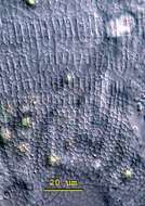
Band-form macronucleus of Arcuospathidum cultriforme scalpriforme (KAHL,1930) FOISSNER,2003.Stained by the methylgreen-pyroninY technique (see Foissner, W.Europ. J. Protistol.27:313-330;1991).Brightfield.
-
Pelagovasicola (pee-ladge-o-vee-sick-o-la) cinctum is a very fast swimming obovoid ciliate measuring 50 - 180 X 40 - 85 microns. It is common in plankton of lakes and ponds. The body is surrounded by 5-7 distinct ciliary girdles. The posterior fifth of the cell is unciliated. The contractile vacuole lies in the posterior end and has about 20 radial collecting channels. The macronucleus is kidney-shaped and lies in the mid-body. Extrusomes are arranged in the margin of the oral dome, occasionally extruded as bundles of fine filaments. This slightly squashed specimen was collected in the plankton of a bog pond near Konstanz, Germany, and this images emphasizes the extruded extrusomes at the margin of the oral dome. Differential interference contrast.
-
Originally described by Schewiakoff under the name Didinium balbianii. The "rear offspring" (i.e. opisthe) of a recent division. cv--Contractile vacuole N--Macronucleus ncl--Micronucleus o--Mouth W--Ciliated ring
-
Lateral view of the haptorid ciliate,Askenasia volvox (Eichwald,1852) Kahl, 1930. The cell is spherical posteriorly and the anterior is a truncate cone.The cytostome is at the anterior apex.The cytostome is surrounded by an undulating line of granules (seen only in silver impregnated specimens).Somatic cilia are arranged in three (anterior,middle and posterior)girdles.The posterior girdle consists of long stiff bristles (seen here).The anterior cilia are directed forward and the middle girdle cilia are longer,curving backwards in a "sickle" configuration.These cilia produce the saltatory locomotion typical of this genus (they are seen well in this image).The posterior of the cell is unciliated.The central macronucleus is C-shaped. There is a single subequatorial contractile vacuole (seen here to viewer's right). From a freshwater pond near Boise, Idaho.Brightfield with closed condenser.
-
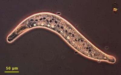
Homalozoon, a elongate ribbon-like predatory ciliate. The body is truncated (cut) off at the front end where the mouth is located, and pointed posteriorly. It has rows of cilia mostly on the ventral side, it glides over the substrate, sometimes contracting. Feeds on detritus and other protists. Phase contrast micrograph.
-
Originally described as Dinophrya lieberkuhnii (Butschli) Shown with the posterior extended to a tail-like appendage. a -- Anus cv -- Contractile vacuole ek -- Ectoplasm N -- Macronucleus nk -- Food particle st -- Cytopharyngeal basket W -- Ciliated ring
-
-
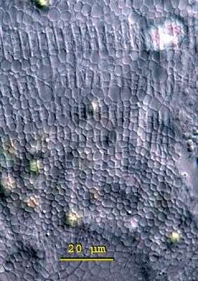
Pelagovasicola (pee-ladge-o-vee-sick-o-la) cinctum is a very fast swimming obovoid ciliate measuring 50 - 180 X 40 - 85 microns. It is common in plankton of lakes and ponds. The body is surrounded by 5-7 distinct ciliary girdles. The posterior fifth of the cell is unciliated. The contractile vacuole lies in the posterior end and has about 20 radial collecting channels. The macronucleus is kidney-shaped and lies in the mid-body. Extrusomes are arranged in the margin of the oral dome, occasionally extruded as bundles of fine filaments. This slightly squashed specimen was collected in the plankton of a bog pond near Konstanz, Germany, and this images emphasizes the finely meshed alveolar pattern of the cortex. Differential interference contrast.
-
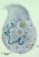
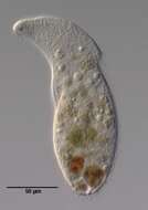
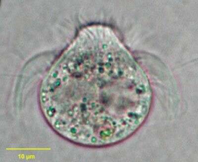
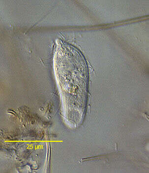
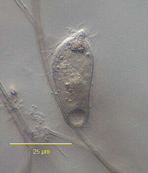
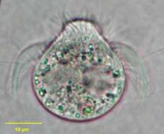
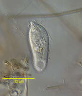
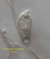
Portrait of Rhopalophrya gracilis (Kahl,1926).Collected from a freshwater pond near Boise, Idaho. DIC.
-
Lateral view of the haptorid ciliate,Askenasia volvox (Eichwald,1852) Kahl, 1930. The cell is spherical posteriorly and the anterior is a truncate cone.The cytostome is at the anterior apex.The cytostome is surrounded by an undulating line of granules (seen only in silver impregnated specimens).Somatic cilia are arranged in three (anterior,middle and posterior)girdles.The posterior girdle consists of long stiff bristles (seen here).The anterior cilia are directed forward and the middle girdle cilia are longer,curving backwards in a "sickle" configuration.These cilia produce the saltatory locomotion typical of this genus (they are seen well in this image).The posterior of the cell is unciliated.The central macronucleus is C-shaped. There is a single subequatorial contractile vacuole (seen here to viewer's right). From a freshwater pond near Boise, Idaho.DIC.
-
Homalozoon, a elongate ribbon-like predatory ciliate. The body is truncated (cut) off at the front end where the mouth is located, and pointed posteriorly. It has rows of cilia mostly on the ventral side, it glides over the substrate, sometimes contracting. Feeds on detritus and other protists. This image is of the mouth and shows the needle-like extrusomes lying under the cell surface - these are used to kill and capture prey. Behind the extrusomes is adense refractile mass that is involved in the feeding process. Differential interference contrast.
-
Originally described as Dinophrya lieberkuhnii (Butschli). Oral view. cl -- Cilia N -- Macronucleus nk -- Fppd particle 0 -- Mouth st -- Cytopharyngeal basket W -- Ciliated ring
-
-
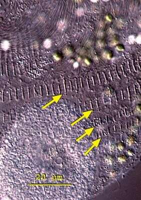
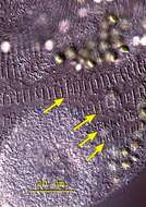
Pelagovasicola (pee-ladge-o-vee-sick-o-la) cinctum is a very fast swimming obovoid ciliate measuring 50 - 180 X 40 - 85 microns. It is common in plankton of lakes and ponds. The body is surrounded by 5-7 distinct ciliary girdles. The posterior fifth of the cell is unciliated. The contractile vacuole lies in the posterior end and has about 20 radial collecting channels. The macronucleus is kidney-shaped and lies in the mid-body. Extrusomes are arranged in the margin of the oral dome, occasionally extruded as bundles of fine filaments. This slightly squashed specimen was collected in the plankton of a bog pond near Konstanz, Germany, and this images emphasizes the characteristic rows of basal bodies ciliary girdles (arrows). Differential interference contrast.
-
Portrait of Rhopalophrya gracilis (Kahl,1926).Collected from a freshwater pond near Boise, Idaho. DIC.
-
Anterolateral view of the infraciliature of the haptorid ciliate,Askenasia volvox (Eichwald,1852) Kahl, 1930. The cell is spherical posteriorly and the anterior is a truncate cone.The cytostome is at the anterior apex.The cytostome is surrounded by an undulating line of granules (seen only in silver impregnated specimens).Somatic kineties are arranged in three (anterior,middle and posterior)girdles seen in this image.The anterior cilia are directed forward and the middle girdle cilia are longer,curving backwards in a "sickle" configuration.These cilia produce the saltatory locomotion typical of this genus.The posterior girdle consists of long stiff bristles.The posterior of the cell is unciliated.The central macronucleus is C-shaped. There is a single subequatorial contractile vacuole.Stained by the silver carbonate technique (see Foissner, W. Europ. J. Protistol., 27:313-330;1991). From a freshwater pond near Boise, Idaho.Brightfield.
-
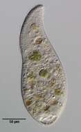
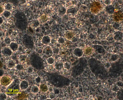
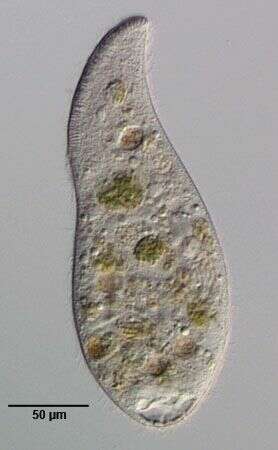
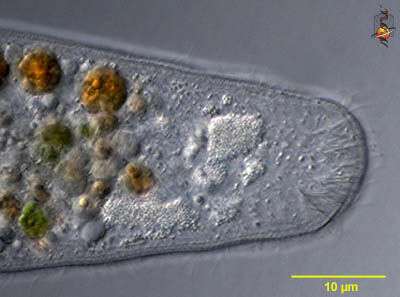
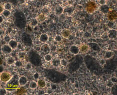
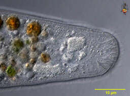
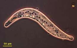
The anterior end is to the left, it is slightly spatulate and contains extrusomes that will key prey organisms - such as other ciliates. The dark inclusions are nodes of the macronucleus and the light regions are contractile vacuoles. There is a large aggregation just behind the mouth and it is believed this contributes membrane to forming food vacuoles. Phase contrast microscopy.