-
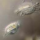
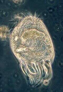
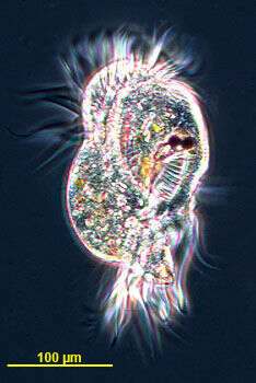
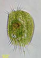
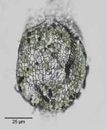
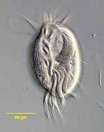
Synopsis of dorsal and ventral view on Euplotes affinis. The species characterizing dorsal ribs together with the adoral zone of membranelles are shown on the left. The ventral cirri (bunched cilia) are used for walking on substrata. Collected from Bodden, the brackish waters lying between the isles of Hiddensee and Ruegen (German Baltic Sea). This image was taken using Zeiss Universal with Olympus C7070 CCD camera.
-
Originally described by Ehrenberg under the name Stylonychia appendiculata.
-
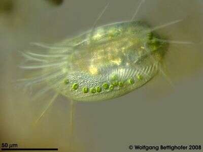
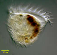
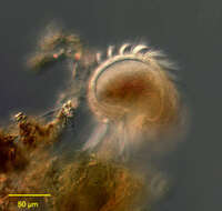
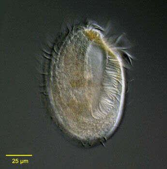
in vivo portrait of Euplotes daidaleos (Diller, Kounaris, 1966) this species in the common and widespread genus of hypotrich ciliates has endosymbiotic green algae. From freshwater pond near Boise, Idaho.Brightfield.
-
trait of the marine hypotrich ciliate, Gastrocirrhus monilifer ((Ozaki et Yagiu, 1942). The cell body is broadly cup-shaped, transversely truncate anteriorly and rounded posteriorly. The broad, funnel-shaped peristome opens anteriorly.The peristome is bordered on the left by a prominent adoral zone of membranelles. There are strongly developed frontoventral and transverse cirri, the latter in a U-shaped distribution posterior to the cytostome. The frontoventral cirri lie to the right of the peristome. There are no caudal cirri.There are a few dorsal kineties with short inconspicuous cilia. The macronucleus is moniliform. Cells are brownish-yellow in color. G. monilifer crawls along the substrate on its cirri. Collected from a commercial saltwater aquarium in Boise, Idaho February 2004. DIC optics.
-
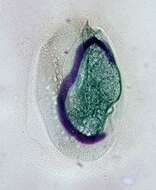
Feulgen stained fixed cell, the stain colours the nuclei purple. The large curving structure is the macronucleus, and a small round micronucleus is located upper left.
-
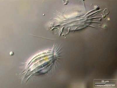
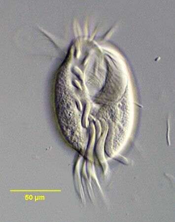
Uronychia is a hypotrich ciliate. As such it has an adoral zone of membranelles for feeding, and these extend around the anterior end of the cell. Like other hypotrichs, motile cilia are clustered into cirri on the ventral surface. Those of Diophrys are very strongly developed. Phase contrast.
-
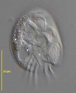
Ventral view of Aspidisca lynceus (MUELLER,1773) EHRENBERG, 1830.Collected from a freshwater irrigation canal in Boise,Idaho December 2007.DIC
-
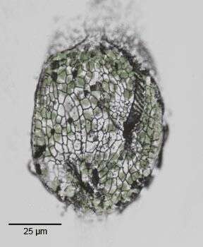
Ventral silverline system of Euplotes daidaleos (Diller, Kounaris, 1966) this species in the common and widespread genus of hypotrich ciliates has endosymbiotic green algae.The adoral zone of membranelles is seen to the viewer's right.The positions of the cirri are indicated by the large black spots.Stained by the dry silver nitrate technique (see Foissner, W. Europ. J. Protistol., 27:313-330;1991) From freshwater pond near Boise Idaho.June 2005.Brightfield.
-
Portrait of the marine hypotrich ciliate, Gastrocirrhus monilifer (Ozaki et Yagiu, 1942). The cell body is broadly cup-shaped, transversely truncate anteriorly and rounded posteriorly. The broad, funnel-shaped peristome opens anteriorly.The peristome is bordered on the left by a prominent adoral zone of membranelles. There are strongly developed frontoventral and transverse cirri, the latter in a U-shaped distribution posterior to the cytostome. The frontoventral cirri lie to the right of the peristome. There are no caudal cirri.There are a few dorsal kineties with short inconspicuous cilia. The macronucleus is moniliform. Cells are brownish-yellow in color. G. monilifer crawls along the substrate on its cirri. Collected from a commercial saltwater aquarium in Boise, Idaho February 2004. DIC optics.
-
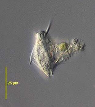
Marine species, very well developed cirri. Phase contrast micrograph of living cell.
-
-
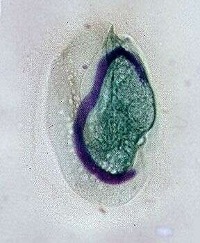
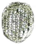
Dorsal silverline system of Euplotes daidaleos (Diller, Kounaris, 1966) this species in the common and widespread genus of hypotrich ciliates has endosymbiotic green algae of the chlorella type (still visible in this stained specimen).The silverline system of this species is the "double-patella" type. Stained by the dry silver nitrate technique (see Foissner, W. Europ. J. Protistol., 27:313-330;1991) From freshwater pond near Boise Idaho.June 2005.Brightfield.
-
Lateral view of the marine hypotrich ciliate, Gastrocirrhus monilifer (Ozaki et Yagiu, 1942).The cell body is broadly cup-shaped, transversely truncate anteriorly and rounded posteriorly. The broad, funnel-shaped peristome opens anteriorly.The peristome is bordered on the left by a prominent adoral zone of membranelles. There are strongly developed frontoventral and transverse cirri, the latter in a U-shaped distribution posterior to the cytostome. The frontoventral cirri lie to the right of the peristome. There are no caudal cirri.There are a few dorsal kineties with short inconspicuous cilia. The macronucleus is moniliform. Cells are brownish-yellow in color. G. monilifer crawls along the substrate on its cirri as seen in this image. Collected from a commercial saltwater aquarium in Boise, Idaho February 2004. DIC optics.
-
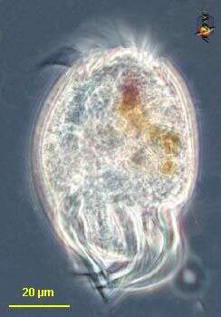
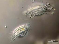
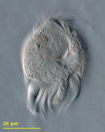
The hypotrich ciliate Uronychia isolated from Little Sippewisset Pond, Woods Hole, MA, USA. Image by Andrew Schurko.
-
Lateral view of the hypotrich ciliate, Aspidisca turrita (Ehrenberg, 1831) Claparède & Lachmann, 1858. The thorn-like dorsal process is clearly seen here (to viewer's left). The first ventral cirrus is also visible projecting anteriorly. Collected from a freshwater pond near Boise, Idaho. June 2005.DIC
-
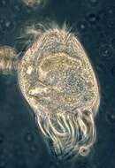
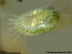
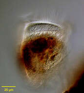
Euplotes daidaleos with its zoochlorellae and ventral cirri. This picture is a digital drawing using 13 frames, manually assembled with Corel Photopaint. Sample from sphagnum pond near Bergenhusen (Schleswig-Holstein, Germany). This image was taken using Zeiss Universal with Olympus C7070 CCD camera.
-
Frontal view of the marine hypotrich ciliate, Gastrocirrhus monilifer (Ozaki et Yagiu, 1942).The cell body is broadly cup-shaped, transversely truncate anteriorly and rounded posteriorly. The broad, funnel-shaped peristome opens anteriorly.The peristome is bordered on the left by a prominent adoral zone of membranelles. There are strongly developed frontoventral and transverse cirri, the latter in a U-shaped distribution posterior to the cytostome. The frontoventral cirri lie to the right of the peristome. There are no caudal cirri.There are a few dorsal kineties with short inconspicuous cilia. The macronucleus is moniliform. Cells are brownish-yellow in color. G. monilifer crawls along the substrate on its cirri as seen in this image. Collected from a commercial saltwater aquarium in Boise, Idaho February 2004. DIC optics.
-
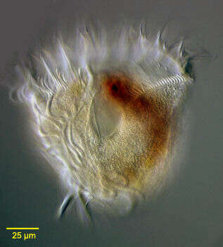
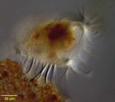
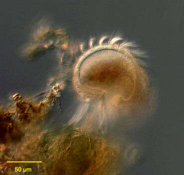
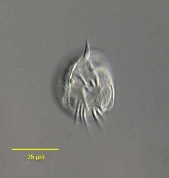
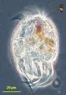
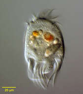
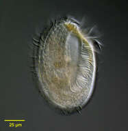
Portrait of the marine hypotrich ciliate, Uronychia transfuga (Müller,1786). Cell outline is ovoid with a colorless rigid pellicle.The broad peristome (not well seen here) is bordered on the left by an adoral zone of membranelles. There are three posterior concavities. Three massive caudal cirri arise from the rightmost concavity. Five transverse cirri arise from the central concavity and three marginal cirri arise from the left concavity (two large and one small). The moniliform macronucleus is arranged in a C-shape.Slow swimming is interrupted by sudden jumping movement. several colorful food vacuoles are visible here. Collected from a commercial saltwater aquarium in Boise, Idaho February 2004. DIC .
-
Ventral view of the hypotrich ciliate, Aspidisca turrita (Ehrenberg, 1831) Claparède & Lachmann, 1858. The cytostome with adoral zone of membranelles is senn at viewer's lower right.The ventral ciiri are seen anteriorly and the transverse cirri posteriorly.Collected from a freshwater pond near Boise, Idaho. June 2005.DIC
-
Detail dorsal view of moniliform macronucleus of the marine hyptorich ciliate, Gastrocirrhus monilifer (Ozaki et Yagiu, 1942). The very similar G. stentoreus has a band-shaped macronucleus.Collected from a commercial saltwater aquarium in Boise, Idaho February 2004. DIC optics.
-
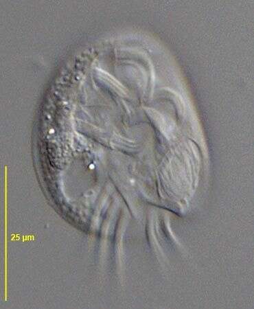
Ventral surface of the hypotricvh ciliate, Uronychia. Isolated from sandy samples taken from Sippiwissett marsh.
-
Originally described by Ehrenberg under the name Euplotes turritus.
-
Portrait (ventral view) of the marine hypotrich ciliate, Kiitricha marina (Nozowa, 1941). The genus is monotypic. The cell has a broadly elliptical outline and is dorsoventrally flattened. The dorsal surface is slightly convex, the ventrum slight concave. The large peristome extends from the anterior end along the left side to terminate in a cytostome in the posterior ¼ of the body. There is a prominent adoral zone of membranelles on the left margin of the peristome and an undulating membrane on its right margin. Uniform dorsal kineties run from left anterior to right posterior on the dorsal surface. On the ventral surface there are approximately 10 longitudinal rows of cirri to the right of and posterior to the cytostome (seen in this image). There are five large transverse cirri lying to the right of the posterior end of the cytostome within the ventral cirral field (seen well in this image). This feature distinguishes Kiitricha marina from the very similar Caryotricha convexa whose transverse cirri lie outside the ventral cirral field posterior and to the left of the peristome. There is a single ovoid macronucleus with an adjacent spherical micronucleus in the center. Collected from a commercial saltwater aquarium in Boise, Idaho February 2004. DIC
-
A species of the marine hypotrich genus, Diophrys (DUJARDIN,1840). The genus contains many species. Collected from a commercial marine aquarium in Boise, Idaho. DIC.