-
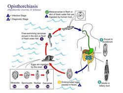
This illustration shows the life cycle of Opisthorchis felineus and O. viverrini, responsible for Opisthorchiasis.Created: 2002
-
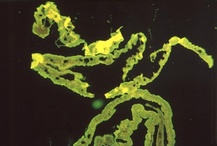
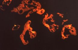
Under a low magnification of 78X, and stained using an indirect fluorescent antibody (IFA) test, this photomicrograph confirmed the presence of Schistosoma mansoni trematodes.Laboratory Diagnosis for Schistosomiasis:Microscopic identification of eggs in stool or urine is the most practical method for diagnosis. Stool examination should be performed when infection with S. mansoni or S. japonicum is suspected, and urine examination should be performed if S. haematobium is suspected. Eggs can be present in the stool in infections with all Schistosoma species. The examination can be performed on a simple smear (1 to 2 mg of fecal material).Created: 1972
-
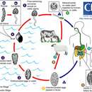
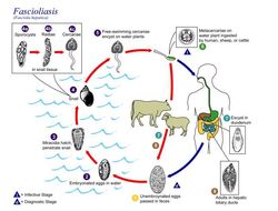
This is an illustration of the life cycle of the causal agents of Fascioliasis.Created: 2002
-
Under a low magnification of 78X, and stained using an indirect fluorescent antibody (IFA) test, this photomicrograph confirmed the presence of Schistosoma mansoni trematodes.Laboratory Diagnosis for Schistosomiasis:Microscopic identification of eggs in stool or urine is the most practical method for diagnosis. Stool examination should be performed when infection with S. mansoni or S. japonicum is suspected, and urine examination should be performed if S. haematobium is suspected. Eggs can be present in the stool in infections with all Schistosoma species. The examination can be performed on a simple smear (1 to 2 mg of fecal material).Created: 1972
-
Under a low magnification of 78X, and stained using an indirect fluorescent antibody (IFA) test, this photomicrograph revealed some of the ultrastructural morphology exhibited by a number of Schistosoma mansoni trematodes.Laboratory Diagnosis for Schistosomiasis:Microscopic identification of eggs in stool or urine is the most practical method for diagnosis. Stool examination should be performed when infection with S. mansoni or S. japonicum is suspected, and urine examination should be performed if S. haematobium is suspected. Eggs can be present in the stool in infections with all Schistosoma species. The examination can be performed on a simple smear (1 to 2 mg of fecal material).Created: 1972
-
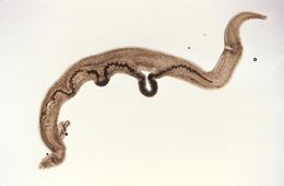
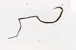
This magnified view reveals a pair of mating Schistosoma mansoni trematodes. Note that the thinner female is cradled inside the thicker male worm's gynecophoral canal.Created: 1973
-
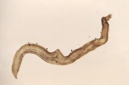
This magnified view reveals a male Schistosoma mansoni trematode. Take a look at PHIL 11193, which depicts a mating pair of worms, where the thinner female is cradled inside the thicker male worm's gynecophoral canal.Created: 1973
-
This magnified view reveals a female Schistosoma mansoni trematode. Take a look at PHIL 11193, which depicts a male S. mansoni, and PHIL 11194, which depicts two mating worms. in which case you can see the thinner female cradled inside the thicker male worm's gynecophoral canal.Created: 1973
-
-
-
-
-
-
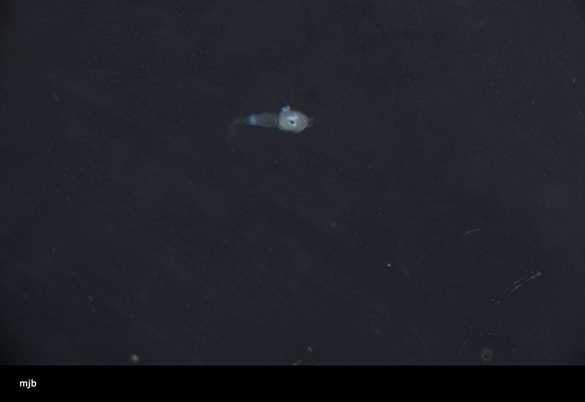
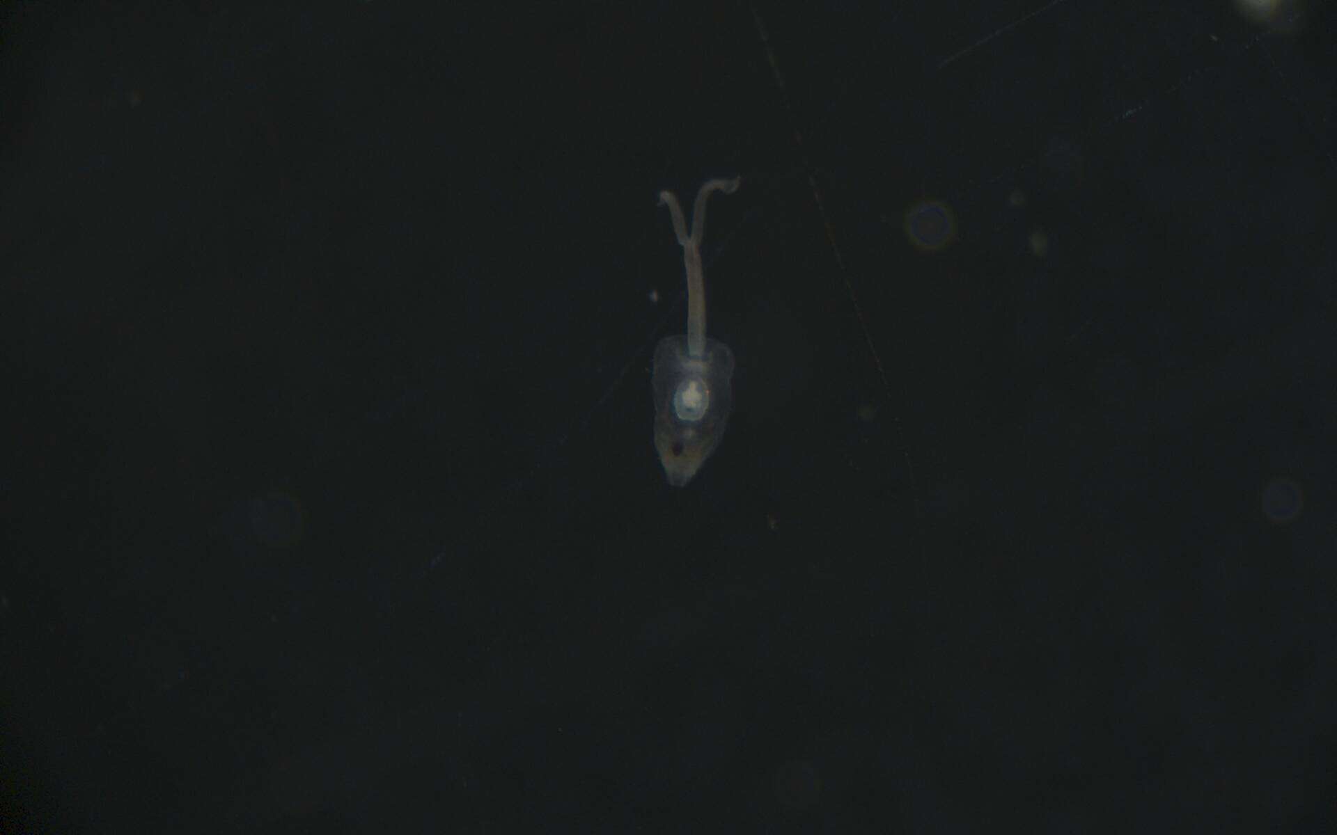
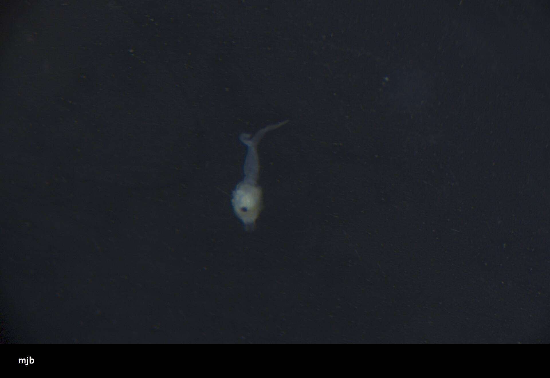
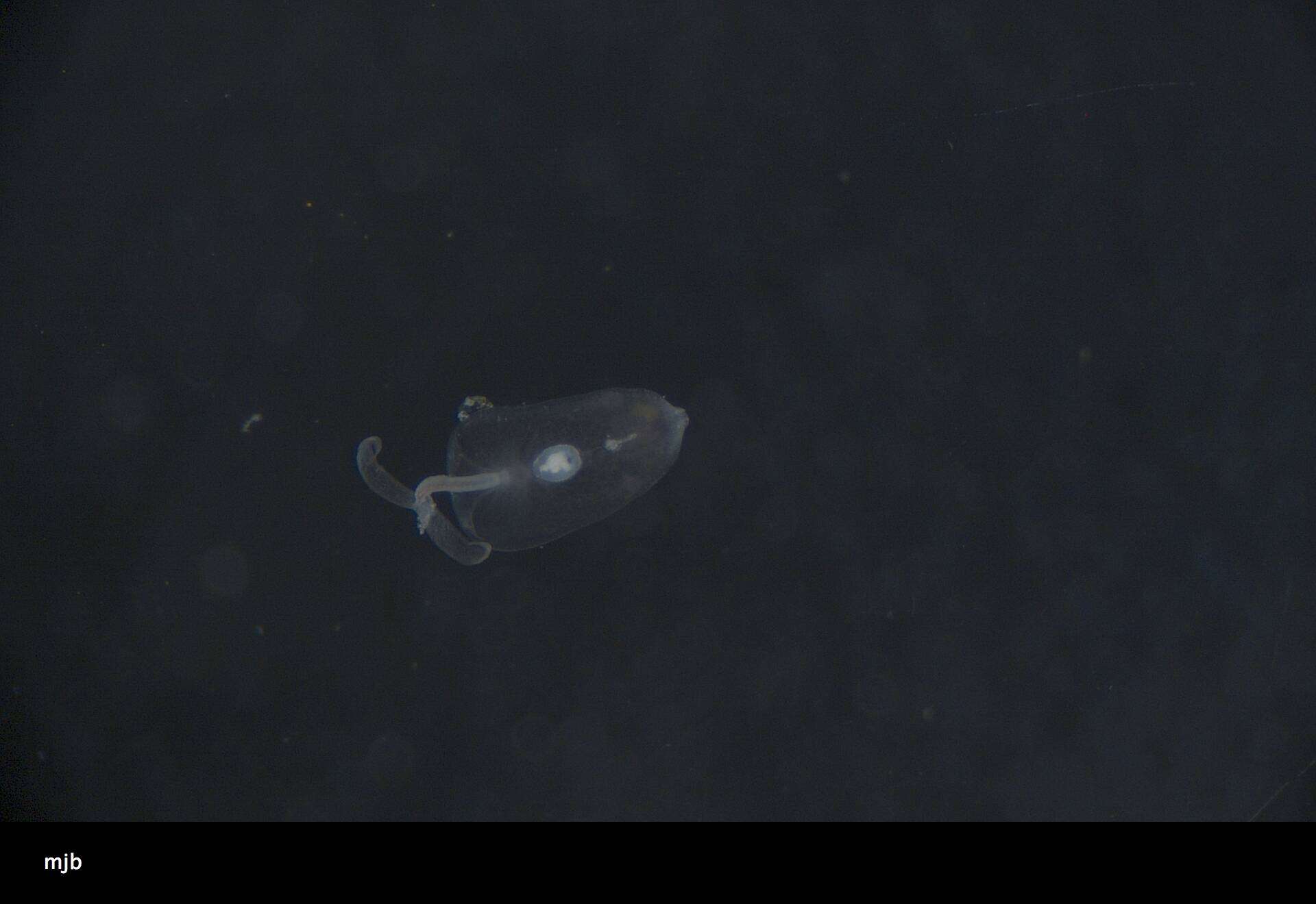
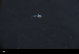
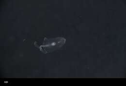
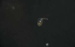
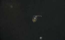
USNM 1617553, FPlrv_510 - Unedited Specimen Image 2. All raw larval photos imported at time of cataloging. Edited, curated photos expected at a later date.
-
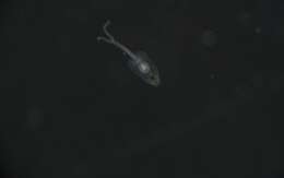
USNM 1618221, FPlrv_1178 - Unedited Specimen Image 1. All raw larval photos imported at time of cataloging. Edited, curated photos expected at a later date.
-
USNM 1617553, FPlrv_510 - Unedited Specimen Image 5. All raw larval photos imported at time of cataloging. Edited, curated photos expected at a later date.
-
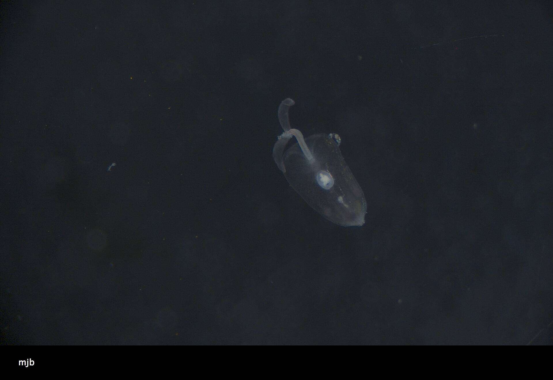
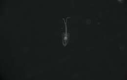
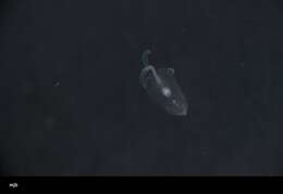
USNM 1617374, FPlrv_331 - Unedited Specimen Image 2. All raw larval photos imported at time of cataloging. Edited, curated photos expected at a later date.
-
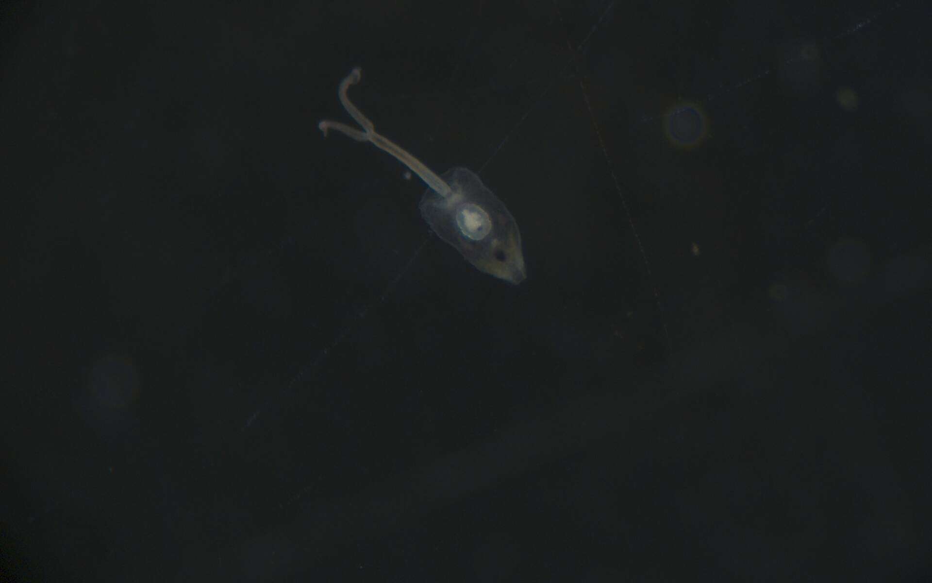
USNM 1617374, FPlrv_331 - Unedited Specimen Image 1. All raw larval photos imported at time of cataloging. Edited, curated photos expected at a later date.
-
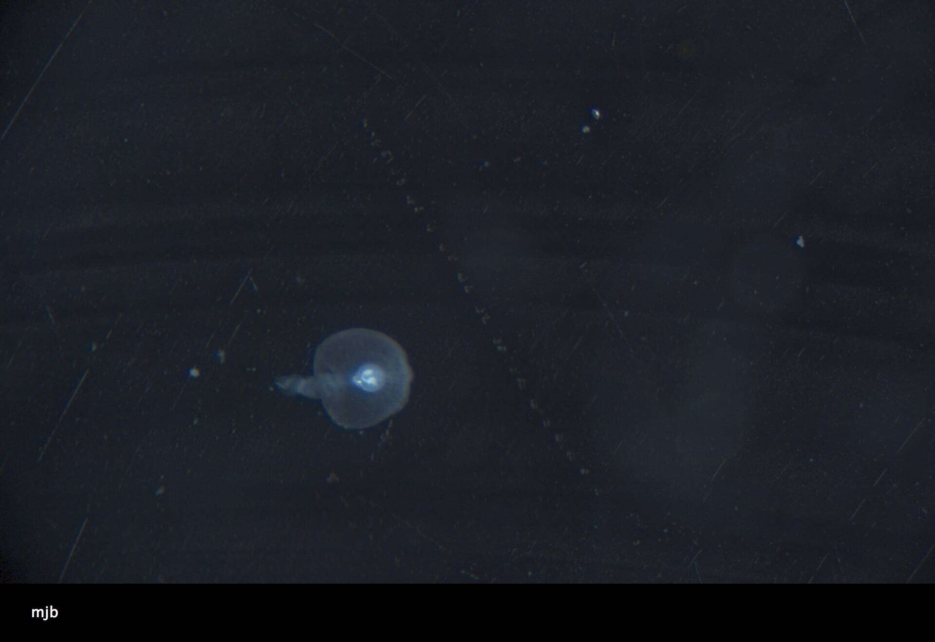
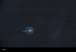
USNM 1617554, FPlrv_511 - Unedited Specimen Image 2. All raw larval photos imported at time of cataloging. Edited, curated photos expected at a later date.
-
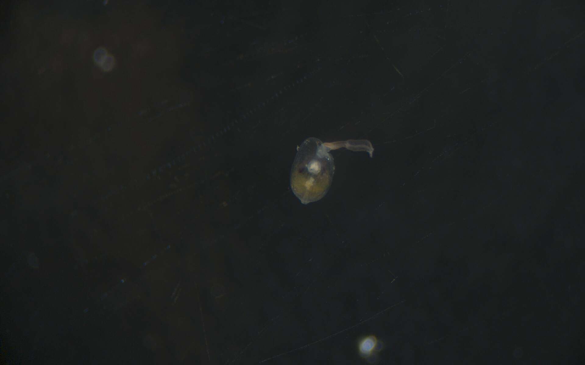
USNM 1617553, FPlrv_510 - Unedited Specimen Image 1. All raw larval photos imported at time of cataloging. Edited, curated photos expected at a later date.
-
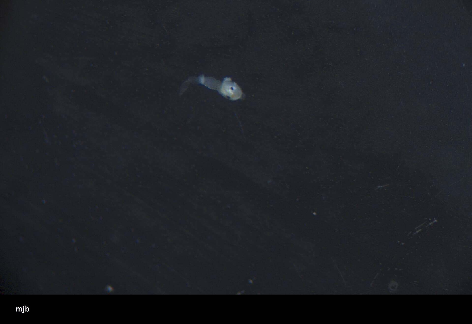
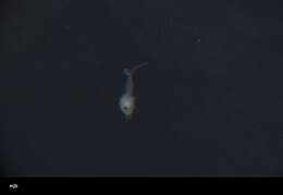
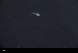
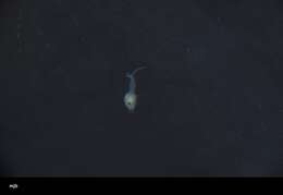
USNM 1618220, FPlrv_1177 - Unedited Specimen Image 3. All raw larval photos imported at time of cataloging. Edited, curated photos expected at a later date.
-
USNM 1618221, FPlrv_1178 - Unedited Specimen Image 3. All raw larval photos imported at time of cataloging. Edited, curated photos expected at a later date.
-
USNM 1618220, FPlrv_1177 - Unedited Specimen Image 2. All raw larval photos imported at time of cataloging. Edited, curated photos expected at a later date.
-
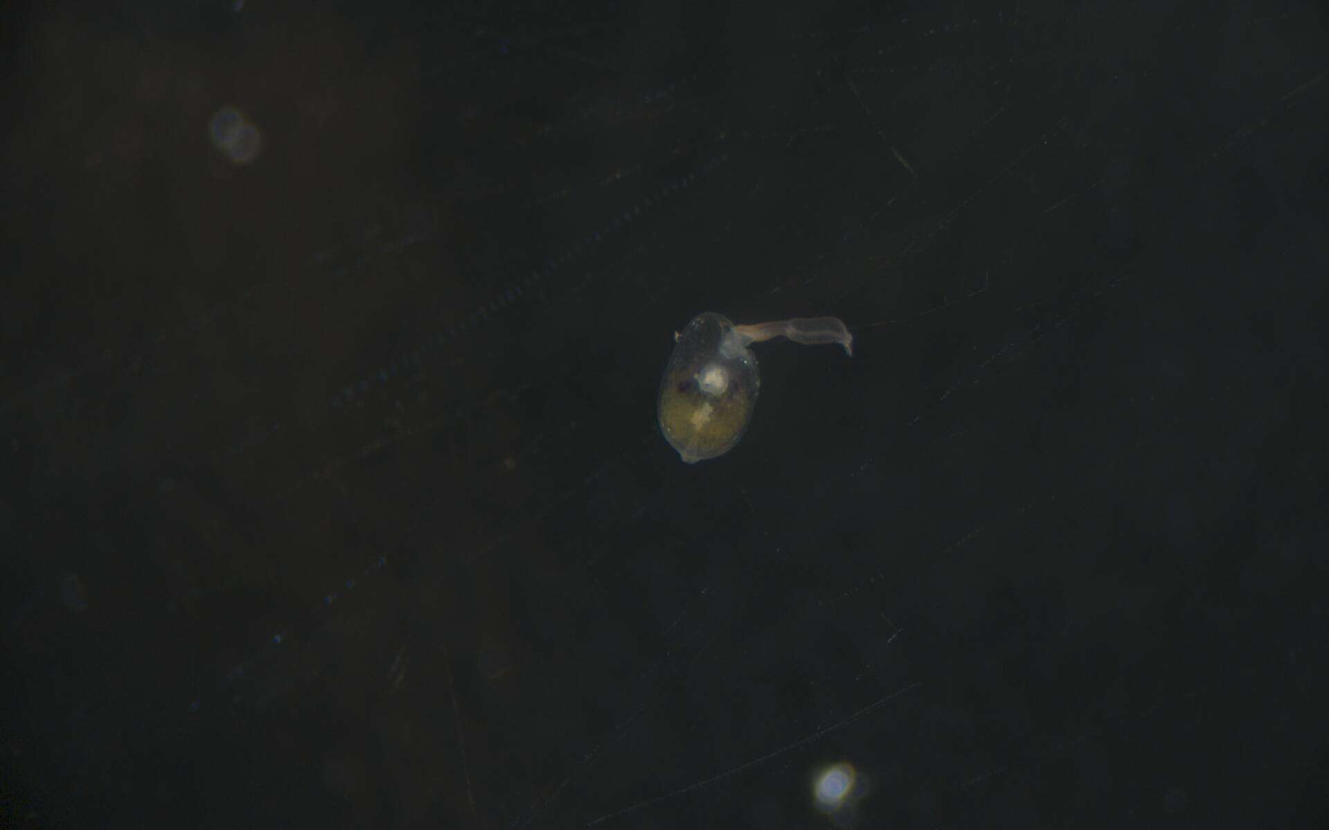
USNM 1617553, FPlrv_510 - Unedited Specimen Image 3. All raw larval photos imported at time of cataloging. Edited, curated photos expected at a later date.