-
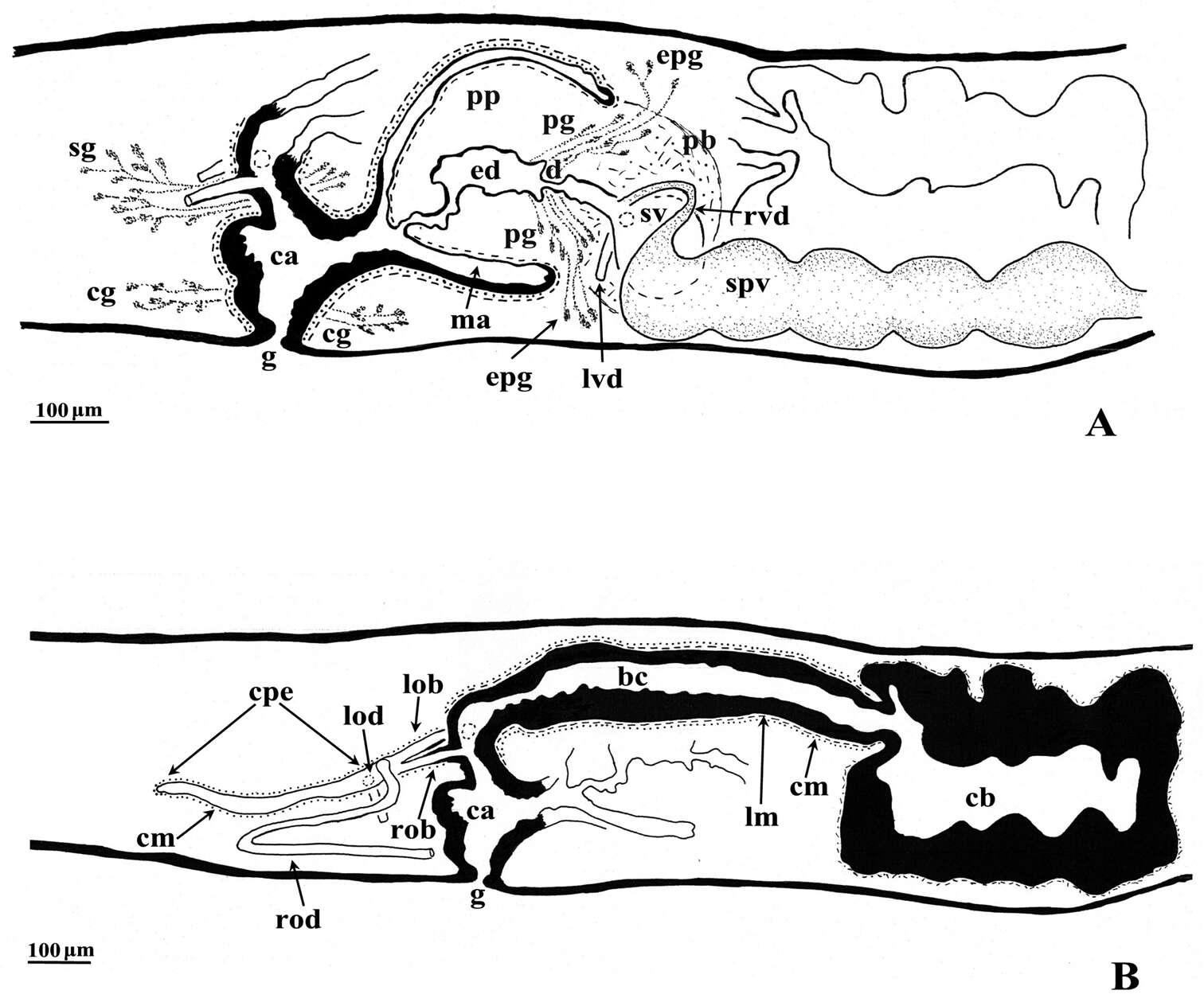
Giacinta Angela Stocchino, Ronald Sluys, Renata Manconi
Zookeys
Figure 3.Dugesia bifida. Holotype ZMA V.Pl. 7189.1, sagittal reconstructions of the copulatory apparatus (anterior to the right), A male copulatory apparatus B female copulatory apparatus.
-
Bothrioplana semperi from Steiermark
-
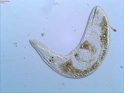
Gyratrix hermaphroditus
-
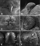
Figure 4.Alaria chengguanensis sp. n., SEM of a female paratype. A Epigyne, ventral view B Epigyne, lateral view C Spinnerets D ALS E PMS F PLS. AC aciniform gland spigot; AG aggregate gland spigot; ALS anterior lateral spinneret; CY cylindrical gland spigot; MAP major ampullate gland spigot; mAP minor ampullate gland spigot; n nubbin; PI piriform gland spigot; PLS posterior lateral spinneret; PMS posterior median spinneret; t tartipore.
-
Giacinta Angela Stocchino, Ronald Sluys, Paolo Deri, Renata Manconi
Zookeys
Figure 4.Dugesia superioris. Photomicrographs of the copulatory apparatus. A Holotype ZMA V.Pl. 7153.1, sagittal section showing the penis bulb and the penis papilla with the ejaculatory duct B Paratype CGAS Pla 6. 3, transverse section of the penis papilla and the ejaculatory duct surrounded by numerous glands C Paratype CGAS Pla 6. 3, transverse section of the bursal canal.
-
Carolina Noreña, Daniel Marquina, Jacinto Perez, Bruno Almon
Zookeys
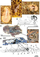
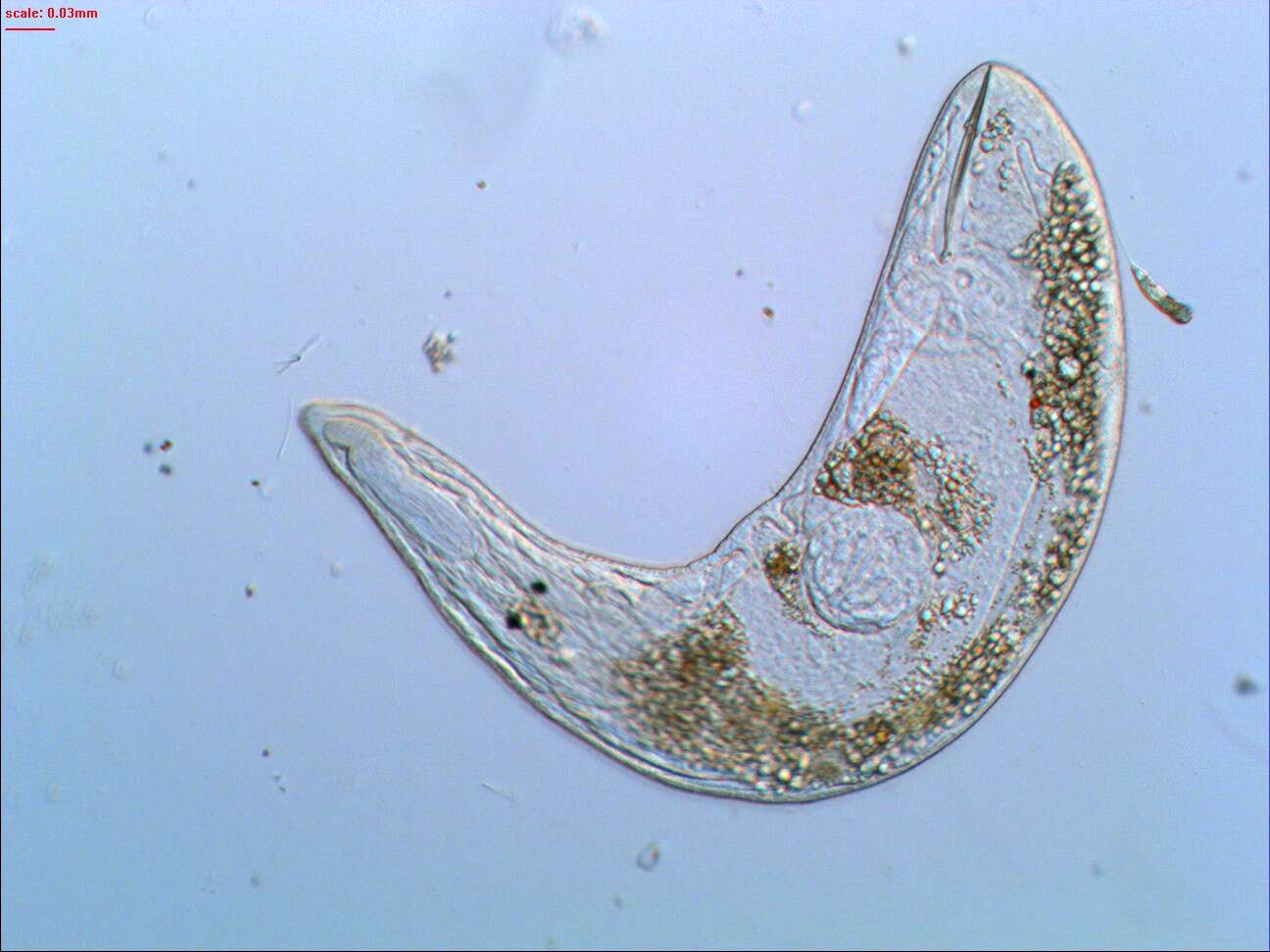
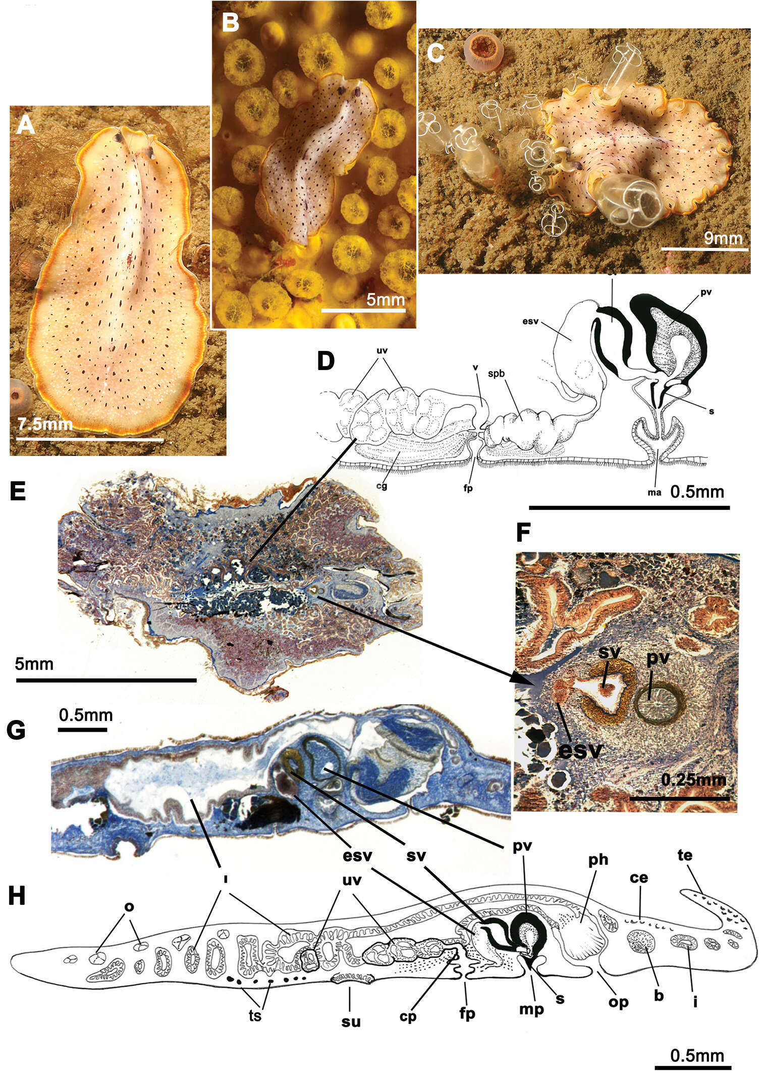
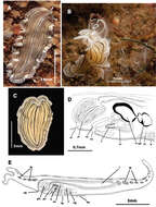
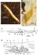
Figure 3.Euryleptodes galikias. A, B dorsal view of a living animal C dorsal and F, ventral views of a fixed specimen E dorsal and D ventral details of the eyes G sagittal reconstruction of a whole specimen H sagittal reconstruction of the reproductive system H. Anterior to the left in A, F and G.
-
Giacinta Angela Stocchino, Ronald Sluys, Renata Manconi
Zookeys
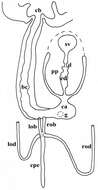
Figure 4.Dugesia bifida. Schematic horizontal reconstruction of the copulatory apparatus.
-
Gyratrix hermaphroditus
-
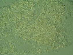
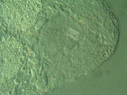
Carcharodopharynx arcanus, a very rare and unusual Dalytyphloplanoid from periodically inundated forest soils near Kloech, Austria.
-
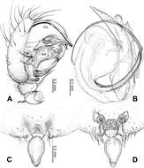
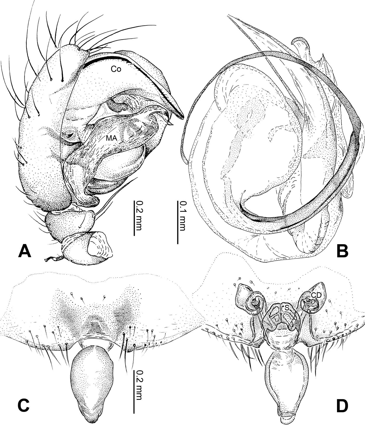
Figure 5.Alaria chengguanensis sp. n., male holotype (A–B) and female paratype (C–D). A Pedipalp, prolateral view B Embolic division, dorsal view C Epigyne, ventral view D Vulva, dorsal view. CD copulatory duct; Co conductor; MA median apophysis; S spermatheca. Scale bars: D as C.
-
Carolina Noreña, Daniel Marquina, Jacinto Perez, Bruno Almon
Zookeys
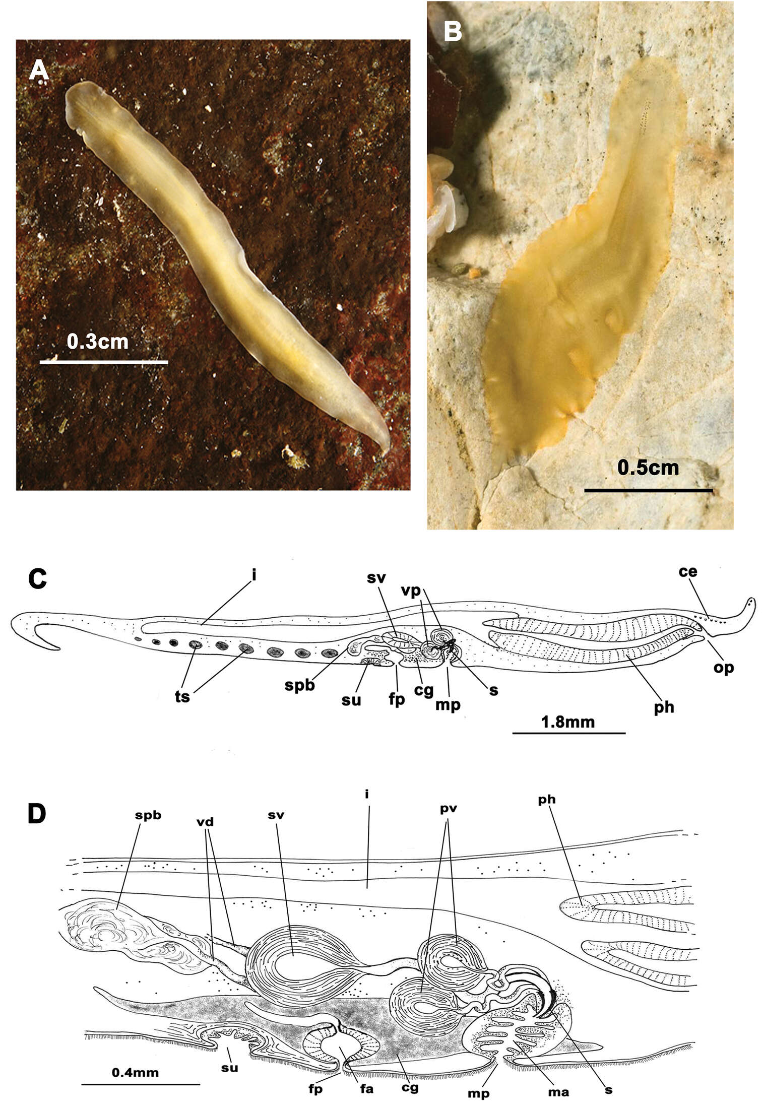
Figure 4.Prostheceraeus vittatus. A dorsal view of a living animal B living animal feeding on ascidians C dorsal view of a fixed specimen D sagittal reconstruction of the copulatory apparatus E sagittal reconstruction of a whole specimen. Anterior to the left in D, E.
-
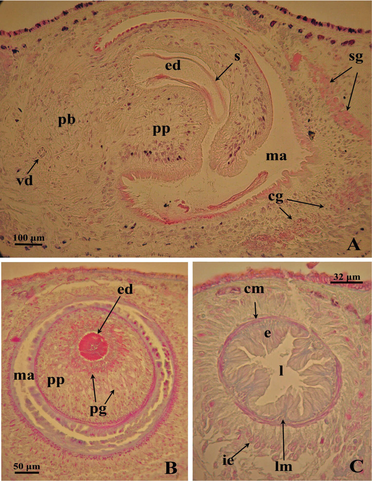
Giacinta Angela Stocchino, Ronald Sluys, Renata Manconi
Zookeys
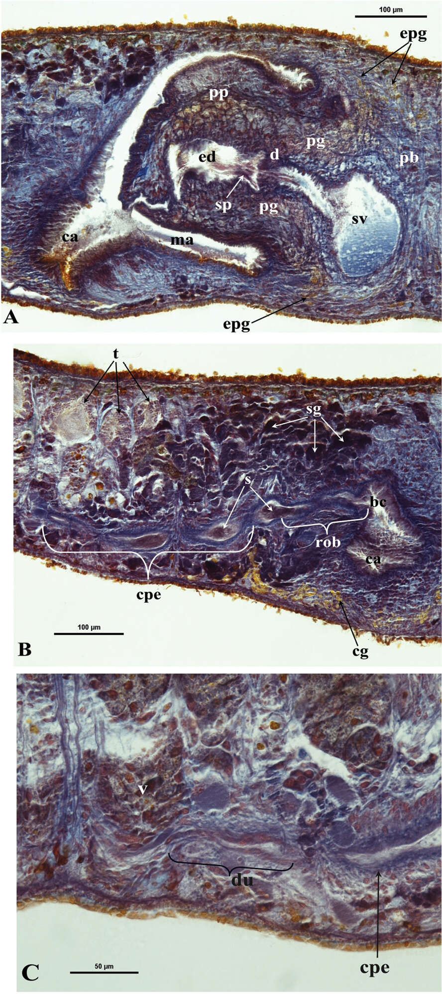
Figure 5.Dugesia bifida. Microphotographs of the copulatory apparatus. A Holotype ZMA V.Pl. 7189.1, sagittal section showing the penis bulb (pb) and the penis papilla (pp) with the seminal vesicle (sv) and the ejaculatory duct (ed) B Holotype ZMA V.Pl. 7189.1, sagittal section showing the opening of the right oviducal branch (rob) through the posterior wall of the bursal canal (bc), and the common posterior oviducal extension (cpe) full of sperm (s) C Paratype CGAS Pla 7.1, sagittal section showing the caudal part of the common posterior oviducal extension (cpe) and the ductule (du) communicating with the ventral part of an adjacent vitellarium (v).
-
Carcharodopharynx arcanus, a very rare and unusual Dalytyphloplanoid from periodically inundated forest soils near Kloech, Austria.
-
Carolina Noreña, Daniel Marquina, Jacinto Perez, Bruno Almon
Zookeys
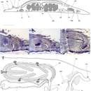
Figure 5.Prostheceraeus moseleyi. A, B, C dorsal views of living animals D sagittal reconstruction of the reproductive system E dorso-ventral histological sections of the whole animal F dorso-ventral histological sections of the copulatory apparatus G sagittal histological section in the region of the pharynx and copulatory apparatus H sagittal reconstruction of a whole specimen. Anterior to the right in C, D, E, F, G and H.
-
Giacinta Angela Stocchino, Ronald Sluys, Renata Manconi
Zookeys
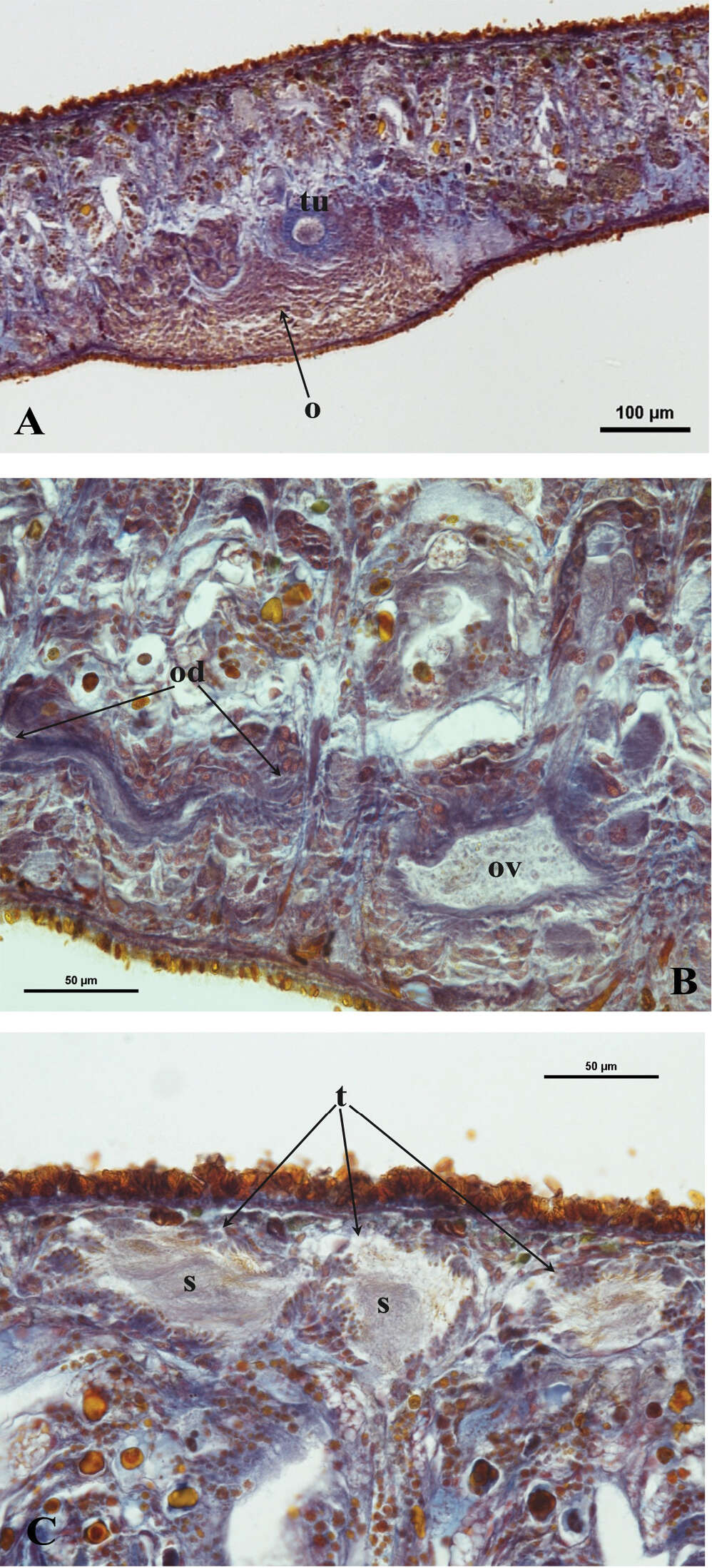
Figure 6.Dugesia bifida. A Holotype ZMA V.Pl. 7189.1, microphotograph of the right hyperplasic ovary (o) with the tuba (tu) B Paratype CGAS Pla 7.1, microphotograph of the oviduct (od) with an expansion (ov) C Holotype ZMA V.Pl. 7189.1, microphotograph of mature testes (t) with sperm (s).
-
Carcharodopharynx arcanus, a very rare and unusual Dalytyphloplanoid from periodically inundated forest soils near Kloech, Austria.
-
Carolina Noreña, Daniel Marquina, Jacinto Perez, Bruno Almon
Zookeys
Figure 6.Prosthiostomum siphunculus. A, B dorsal views of living animals C sagittal reconstruction of a whole specimen D sagittal reconstruction of the copulatory apparatus. Anterior to the right in figures C, D.
-
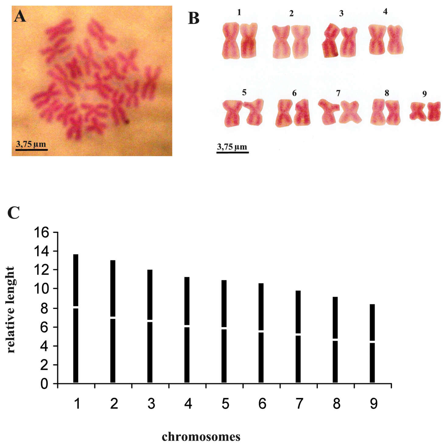
Giacinta Angela Stocchino, Ronald Sluys, Renata Manconi
Zookeys
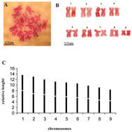
Figure 7.Dugesia bifida. A metaphasic plate B karyogram C idiogram.
-
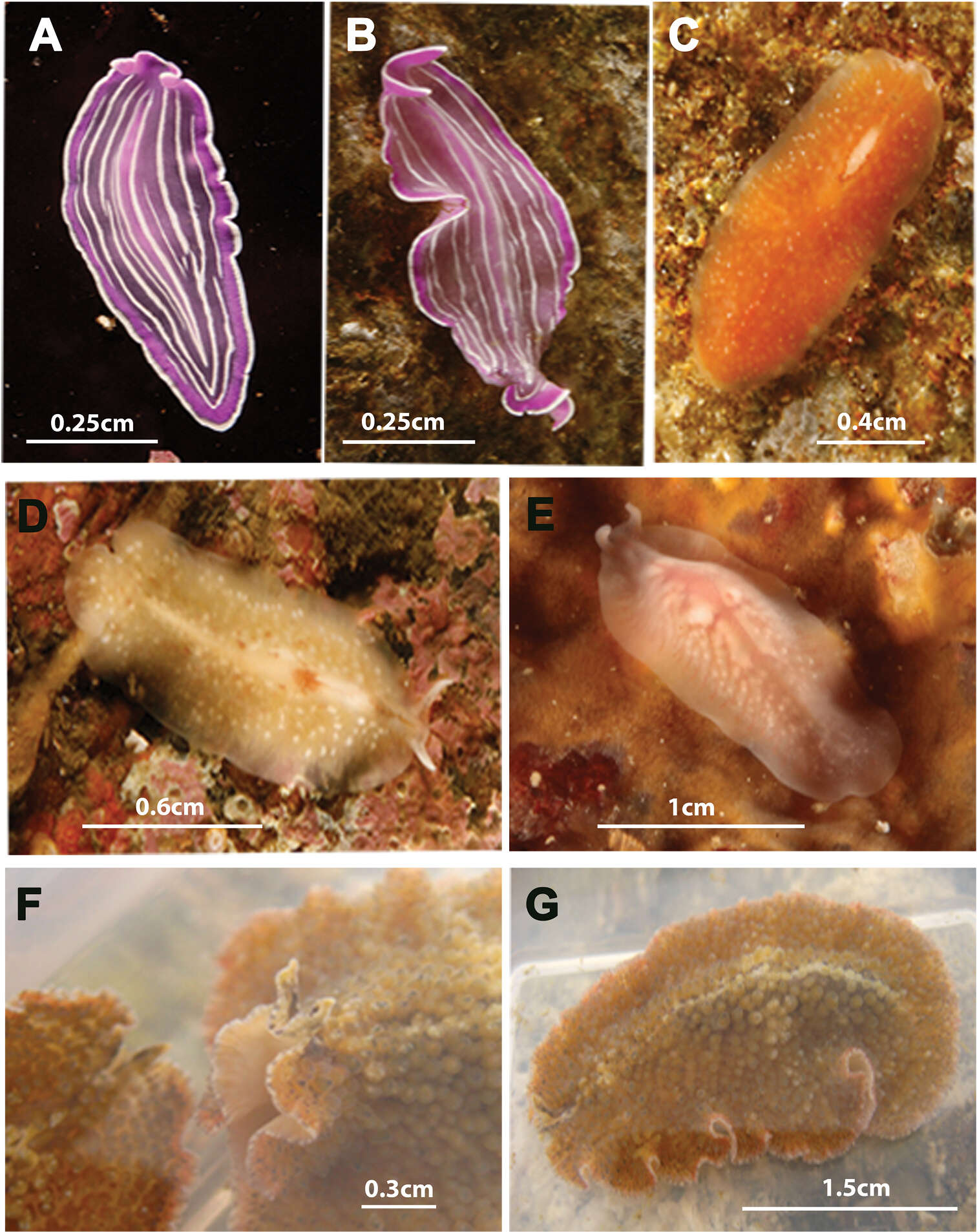
Carolina Noreña, Daniel Marquina, Jacinto Perez, Bruno Almon
Zookeys
Figure 7.Species photographed but not collected. A, B Prostheceraeus roseus C Stylostomum ellipse D, E Oligocladus sanguinolentus F, G Thysanozoon brocchii.
-
Carolina Noreña, Daniel Marquina, Jacinto Perez, Bruno Almon
Zookeys
Figure 7.Species photographed but not collected. A, B Prostheceraeus roseus C Stylostomum ellipse D, E Oligocladus sanguinolentus F, G Thysanozoon brocchii.
-
Carolina Noreña, Daniel Marquina, Jacinto Perez, Bruno Almon
Zookeys
Figure 7.Species photographed but not collected. A, B Prostheceraeus roseus C Stylostomum ellipse D, E Oligocladus sanguinolentus F, G Thysanozoon brocchii.
-
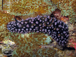
Acoela (small reddish disk shapes) on a Phyllidia Nudibranch
-
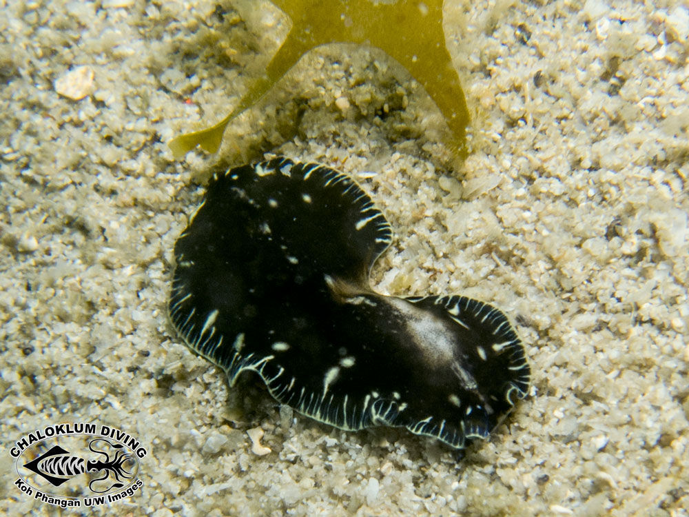
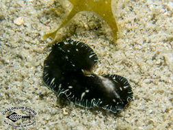
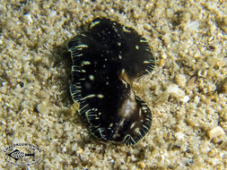
Eurylepta species (damaged)
-
Eurylepta species (damaged)