-
Hobro, Jylland, Danmark
-
Centers for Disease Control/Division of Parasitic Diseases and Malaria
EOL staff
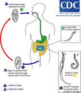
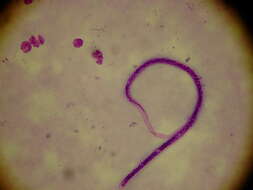
Life cycle of the Human Pinworm (Enterobius vermicularis and V. gregorii)Human pinworm eggs are deposited on the human host's perianal folds (1). Self-infection occurs by transferring infective eggs to the mouth with hands that have scratched the perianal area (2). Person-to-person transmission can also occur through handling of contaminated clothes or bed linens. Enterobiasis (pinworm infection) may also be acquired from surfaces in the environment that are contaminated with pinworm eggs (e.g., curtains, carpeting). A small number of eggs may become airborne and inhaled. These would be swallowed and follow the same development as ingested eggs. Following ingestion of infective eggs, the larvae hatch in the small intestine (3) and the adults establish themselves in the colon (4). The time interval from ingestion of infective eggs to oviposition by the adult females is about one month. The life span of the adults is about two months. Gravid females migrate nocturnally outside the anus and oviposit while crawling on the skin of the perianal area (5). The larvae contained inside the eggs develop (and the eggs become infective) in 4 to 6 hours under optimal conditions (1). Retroinfection, or the migration of newly hatched larvae from the anal skin back into the rectum, may occur but the frequency with which this happens is unknown.From
Centers for Disease Control Parasites and Health website.
-
Saša Širca, Gregor Urek, Stela Lazarova, Milka Elshishka, Vlada Peneva
Zookeys
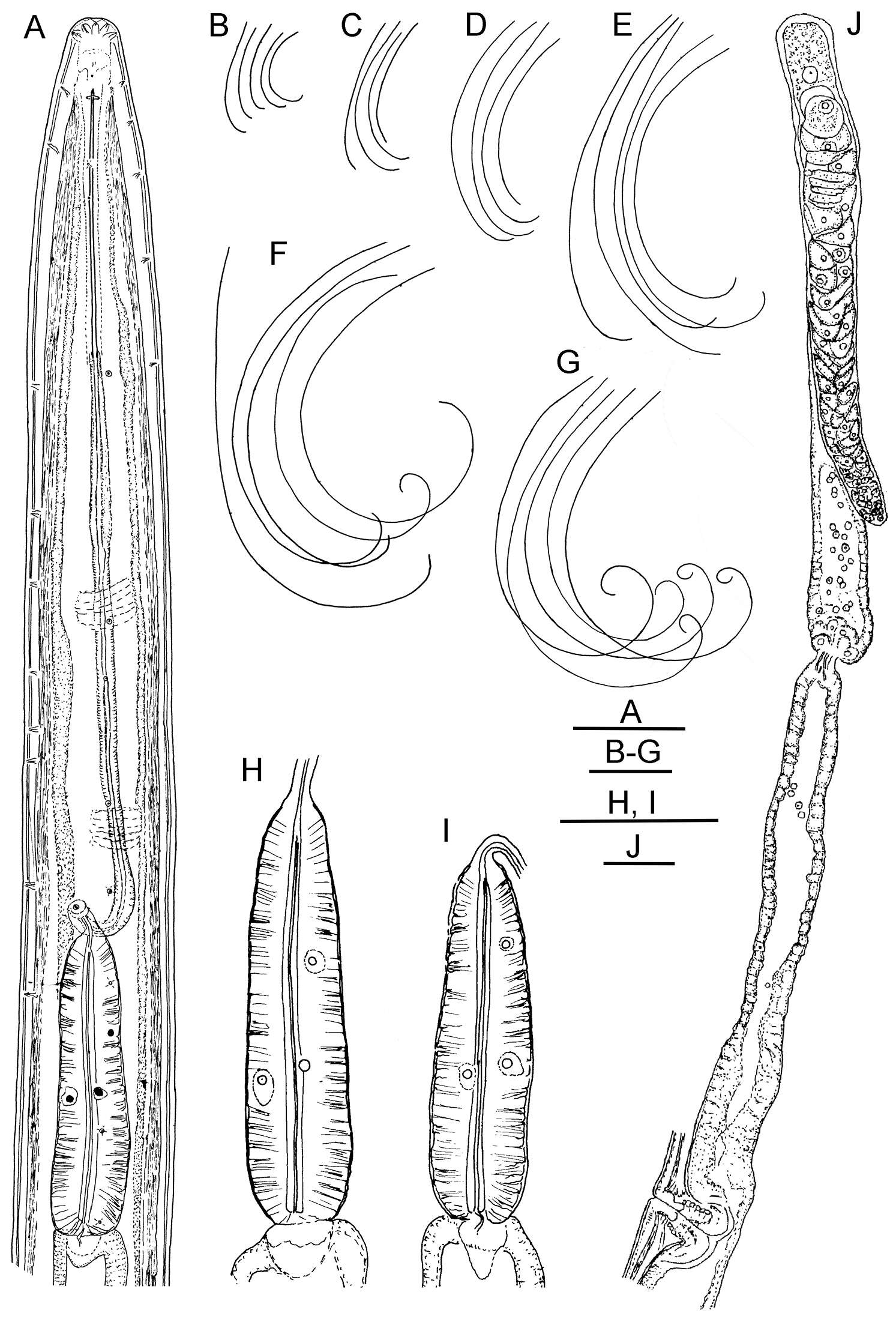
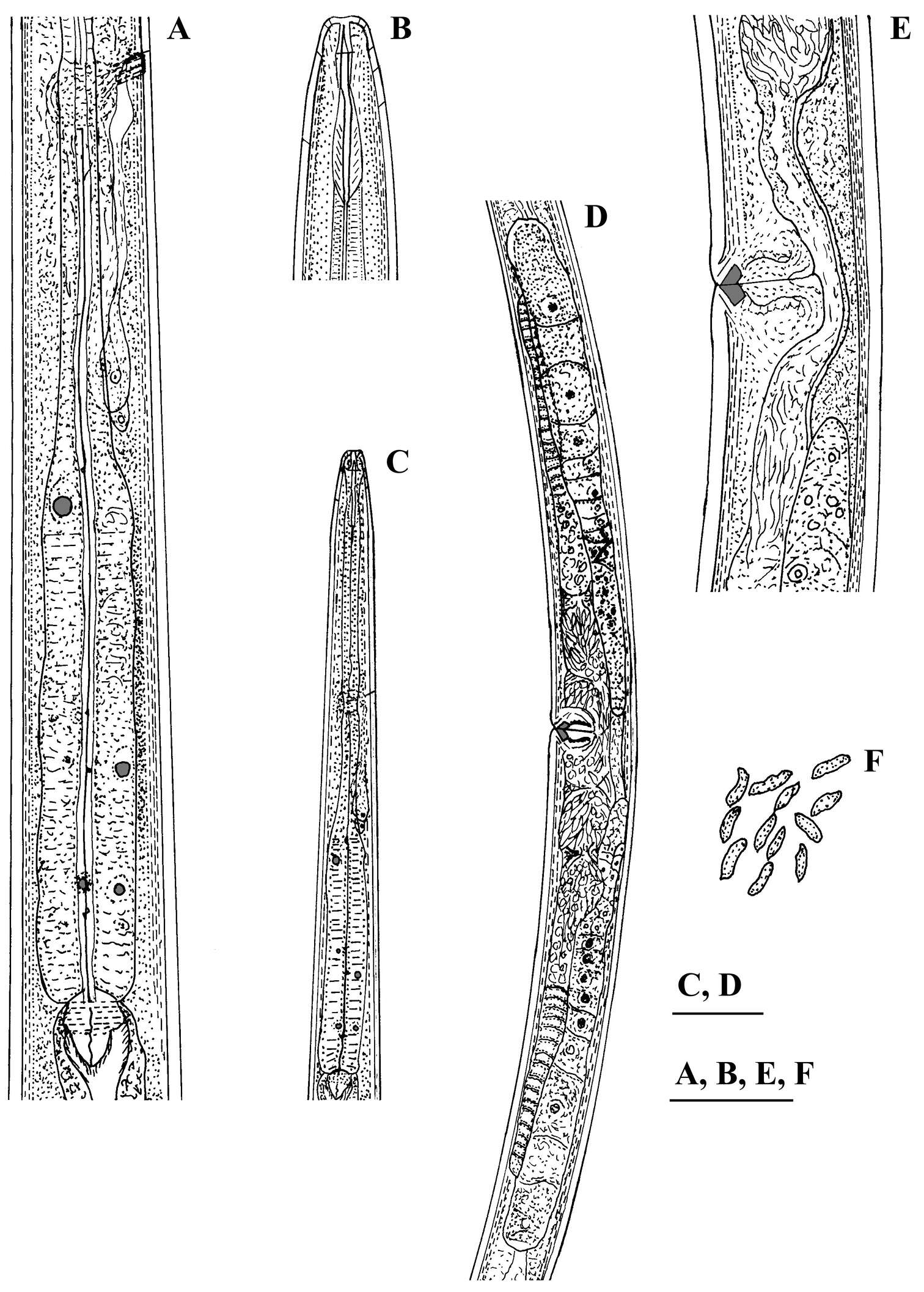
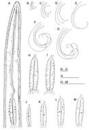
Figure 1.Longidorus carniolensis sp. n. Female: A Neck region F Habitus H Pharyngeal bulb Male: G Habitus I Pharyngeal bulb; Juveniles: B–E Habitus of first, second, third and forth juvenile stages J–M Pharyngeal bulb of first, second, third and forth juvenile stages. Scale bars: B–G 1 mm; A, H–M 100 μm.
-
Habibeh Jabbari, Gholamreza Niknam, Maria Teresa Vinciguerra, Shalaleh Moslehi, Joaquín Abolafia, Reyes Peña-Santiago
Zookeys
Figure 1.Crassolabium persicum sp. n. (all images are in lateral view) A Anterior region B Lip region and amphid fovea in surface C Pharyngeal expansion D Vagina E Spicules and lateral guiding piece F Female, posterior body region G Female, anterior genital branch H Male, posterior body region I Male, entire J Female, entire.
-
Vlada K. Peneva, Stela S. Lazarova, Francesca De Luca, Derek J. F. Brown
Zookeys
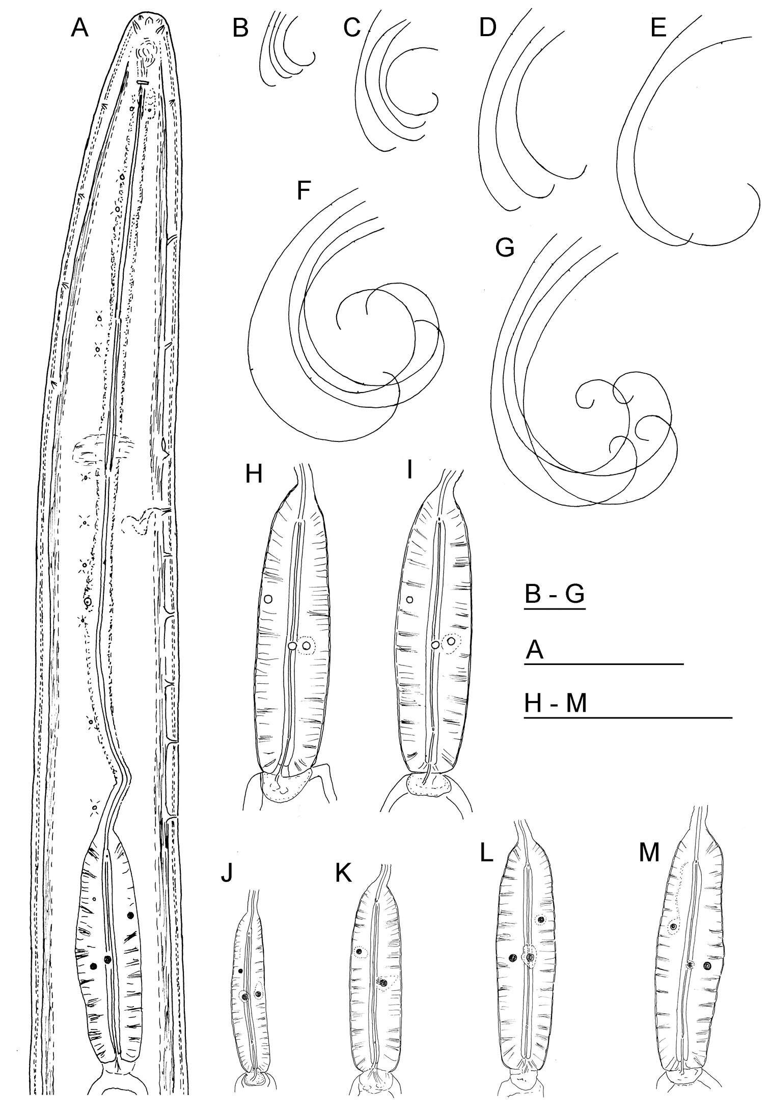
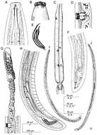
Figure 1.Longidorus cholevae sp. n. Female: A Anterior end F Habitus I Pharyngeal bulb J Anterior genital branch Male: G Habitus H Pharyngeal bulb Juveniles: B–E Habitus of first-, second-, third- and fourth-stage juveniles. Scale-bars: A, H, I, J 50 μm; B–G 1 mm.
-
Sevdan Nedelchev, Milka Elshishka, Stela Lazarova, Georgi Radoslavov, Peter Hristov, Vlada Peneva
Zookeys
Figure 2.Calcaridorylaimus castaneae sp. n. Female: A Pharyngeal gland nuclei B Anterior region C Pharyngeal region D Genital system E Vulval region F Sperm cells in uterus. Scale bars: A, B, E, F – 30 μm; C, D – 50 μm.
-
Figure 2.Longior longior Morffe & García sp. n. (female). A Esophageal region B Cephalic end C Tail, ventral view D Vulva E Egg F Genital tract G Entire nematode, lateral view.
-
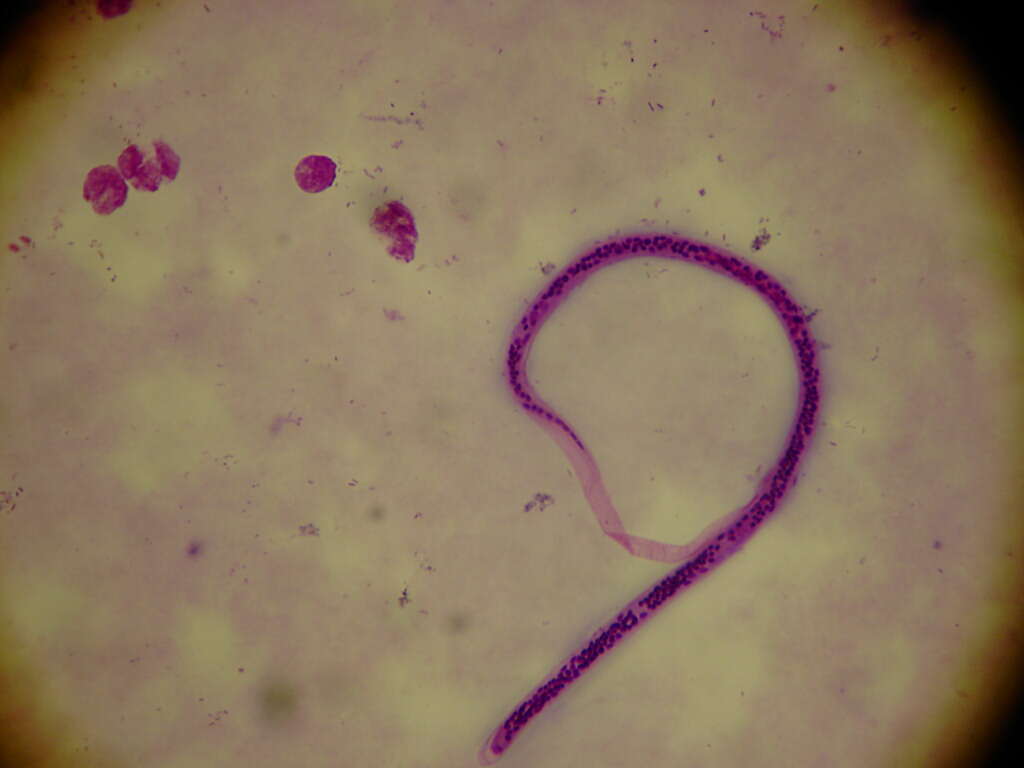
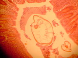
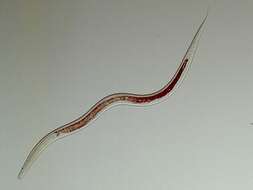
Enterobius vermicularis
-
Wuchereria bancrofti
-
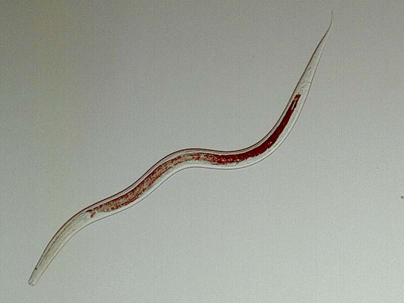
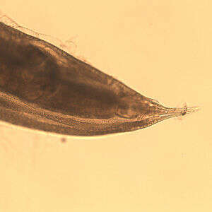
Trichinella spiralis
-
Genus Monhystera body is mostly tapering considerably posteriorly. Caudal sucker small, somewhat pointed. Setae very few. Pharyngeal cavity none. Oesophagus uniform. Spicules long and narrow. Ocellus single, often absent.
-
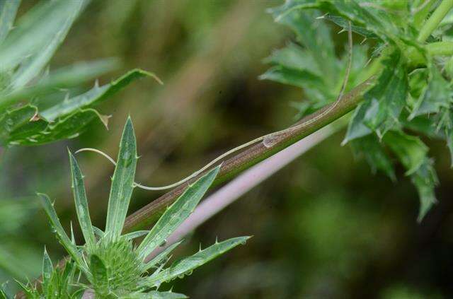
Tent Rock National Monument, NM
-
Hobro, Jylland, Danmark
-
Öesophagostomum sp. adult femalePosterior end of a female Oesophagostomum sp., showing the pointed tail.From
CDC DPDx website
-
Saša Širca, Gregor Urek, Stela Lazarova, Milka Elshishka, Vlada Peneva
Zookeys
Figure 10.Longidorus carniolensis sp. n. Juvenile: A–D Anterior region of first, second, third and forth stages H–K Pharyngeal bulb of first, second, third and forth juvenile stages M, F, G, R genital primordium of first, second, third and forth stages N, S Tail shape of first stage O, T Tail shape of second stage P, U Tail shape of third stage Q, V Tail shape of forth stage Female: E Anterior region L Pharyngeal bulb W Tail shape. Scale bar: 50 μm.
-
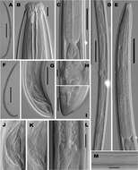
Habibeh Jabbari, Gholamreza Niknam, Maria Teresa Vinciguerra, Shalaleh Moslehi, Joaquín Abolafia, Reyes Peña-Santiago
Zookeys
Figure 2.Crassolabium persicum sp. n. (light micrographs all in lateral view).A Female, entire B Anterior region C Pharyngo–intestinal junction D Female, genital system E Neck region F Male, entire G Male, posterior region H Vagina I Female, caudal region J Spicules K Lateral guiding piece L Oviduct–uterus junction M Lateral chord and pores. (Scale bars: A, F – 500 µm; B, H, K – 10 µm; C, G – 50 µm; D, E – 100 µm; I, J, L, M – 20 µm).
-
Vlada K. Peneva, Stela S. Lazarova, Francesca De Luca, Derek J. F. Brown
Zookeys
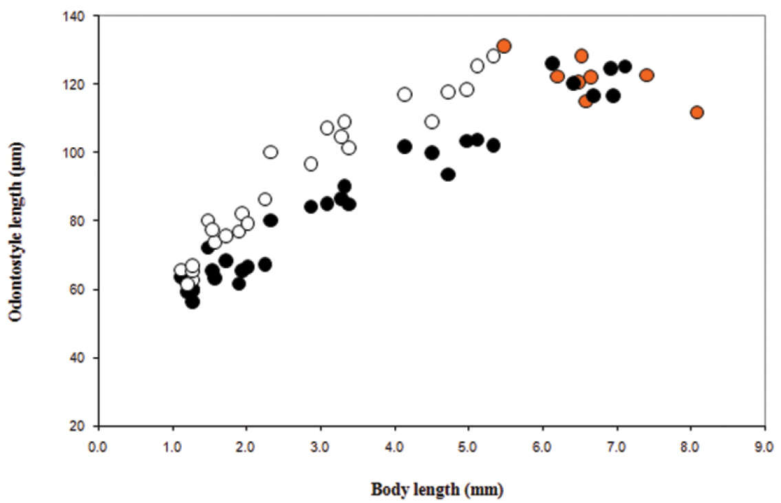
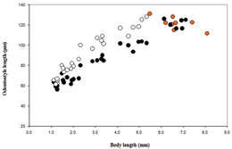
Figure 9.Longidorus cholevae sp. n. Scatter plot of the functional (˜, juveniles and adults, females in orange) and replacement (™, juveniles) odontostyle in relation to body length of the juvenile developmental stages and adults.
-
Sevdan Nedelchev, Milka Elshishka, Stela Lazarova, Georgi Radoslavov, Peter Hristov, Vlada Peneva
Zookeys
Figure 3.Calcaridorylaimus castaneae sp. n. Female: A Posterior region. Male: B, C Extruded spicules with supplements D Posterior region. Scale bar: A–D – 30 μm.
-
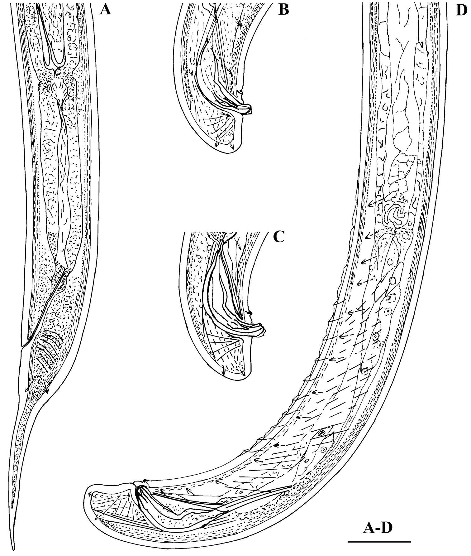
Figure 3.Longior longior Morffe & Garcíasp. n. (male). A Entire nematode, lateral view B Esophageal region, lateral view C Cephalic end D Tail, lateral view.
-
Enterobius vermicularis
-
Hobro, Jylland, Danmark
-
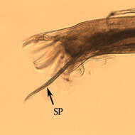
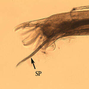
Male Oesophagostomum sp. Posterior end of male Oesophagostomum sp. showing the bursa. Note the spicule (SP).From
CDC DPDx Website
-
Saša Širca, Gregor Urek, Stela Lazarova, Milka Elshishka, Vlada Peneva
Zookeys
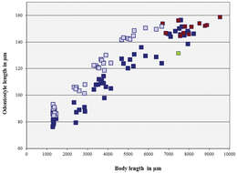
Figure 11.Longidorus carniolensis sp. n. Scatter plot of the functional (, dark blue) and replacement odontostyle (, light blue) in relation to the body length of the juvenile stages and adults: females (, dark blue) and males (, red), female with very short odontostyle (, green).
-
Vlada K. Peneva, Stela S. Lazarova, Francesca De Luca, Derek J. F. Brown
Zookeys
Figure 10.Phylogenetic relationships of Longidorus cholevae sp. n. and its closest species for the D2-D3 rDNA. Bayesian Inference strict consensus tree acquired under GTR+G model. Numbers at the nodes indicating posterior probabilities higher that 0.8 and bootstrap values more that 70% for ML and NJ are presented.