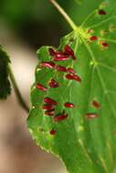
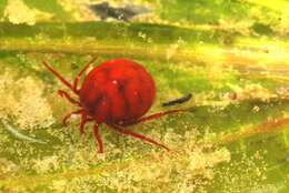
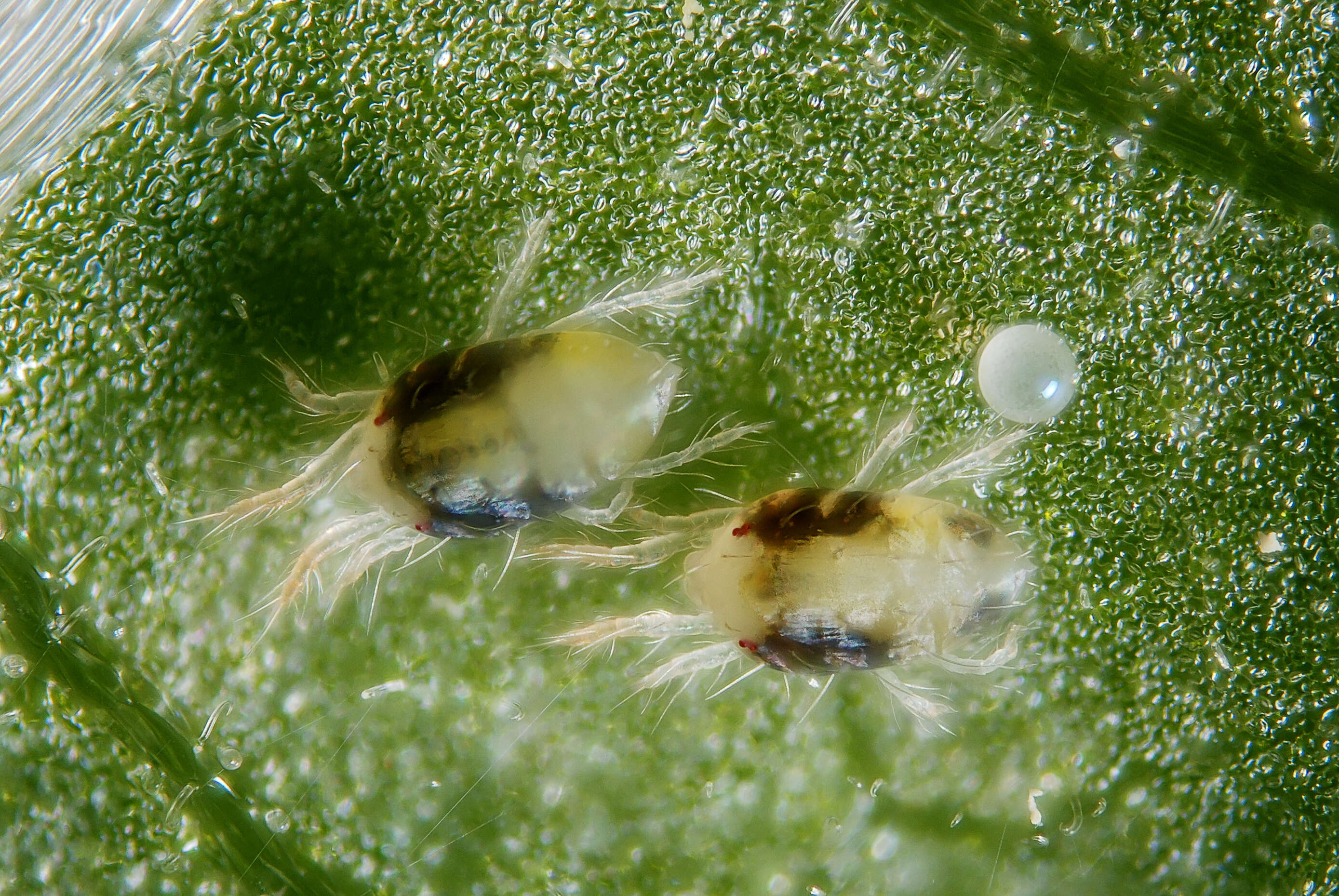
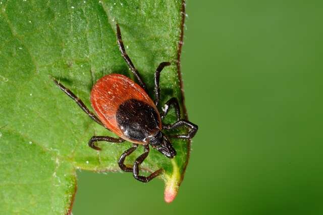
Two females with an egg of the green form of the spider mite Tetranychus urticae.Scale : mite body length ~0.5 mmTechnical settings : - focus stack of 11 images- microscope objective (Nikon achromatic 10x 160/0.25) on bellow (70 mm extention)
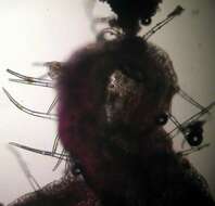
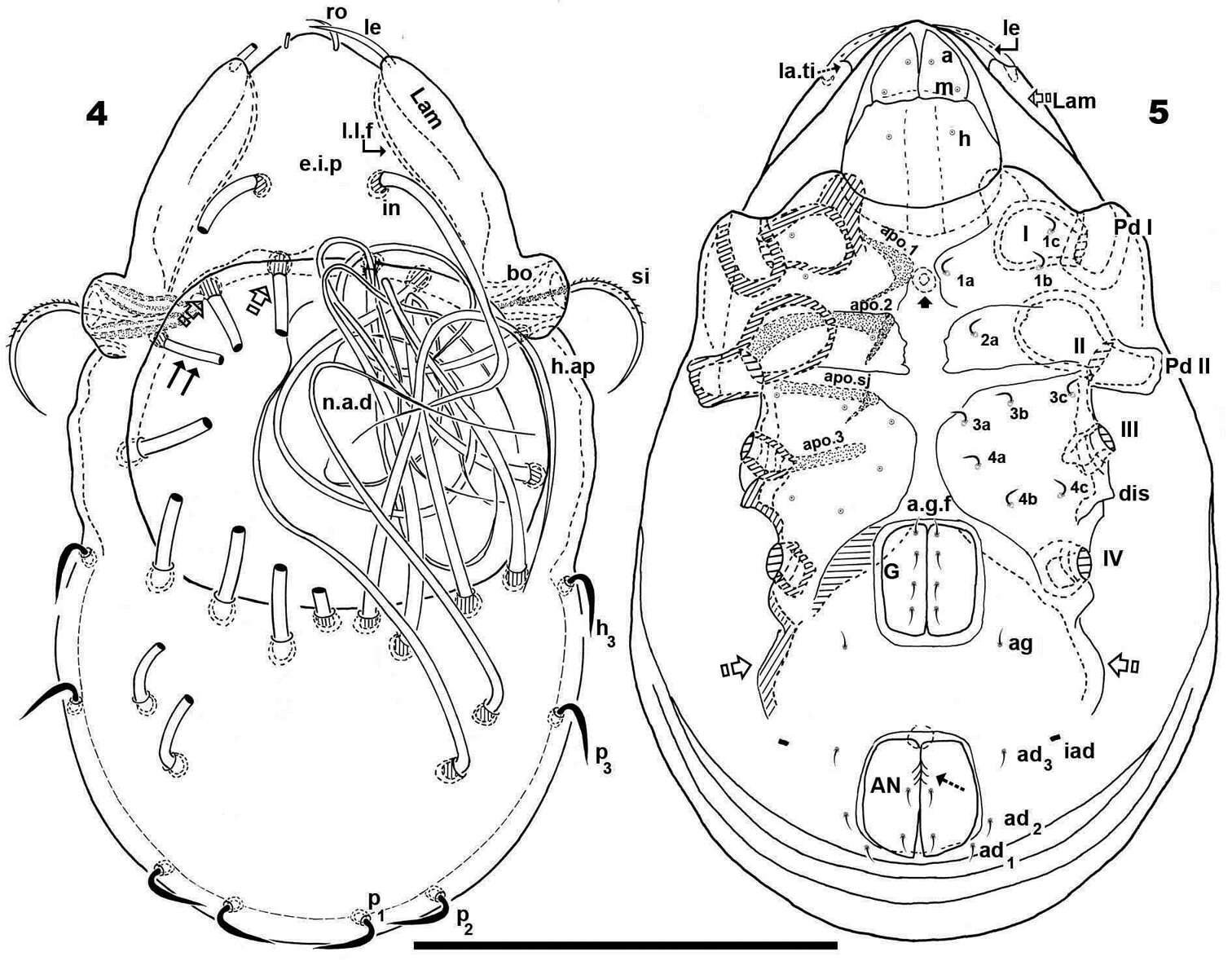
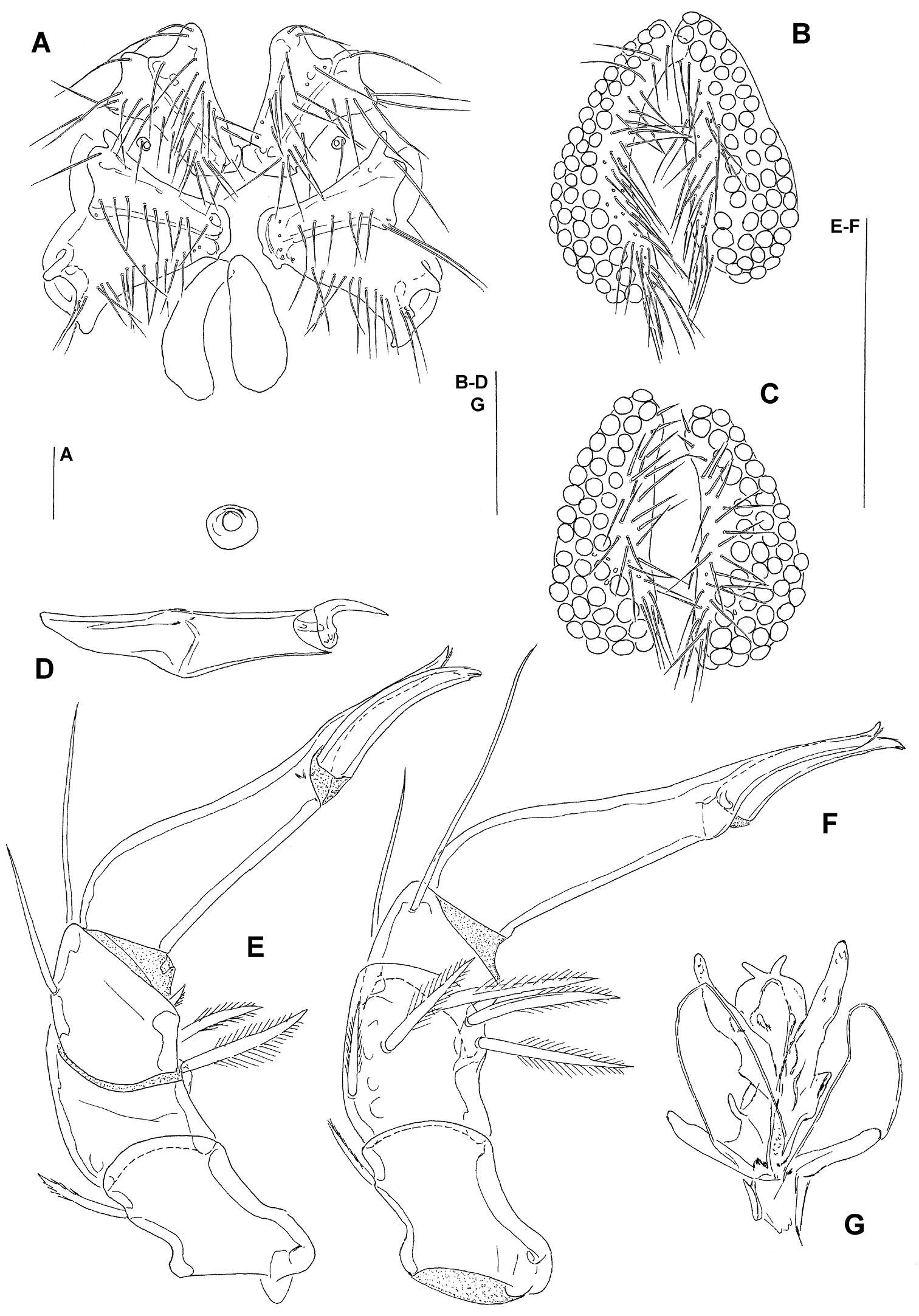
Figure 1. A–G Hydrodroma meridionalis sp.n. (A–B, D, G = male holotype, C, E, F = female paratype) A = coxal and genital field B–C = genital field D = chelicera E = palp, lateral view F = palp, medial view G = ejaculatory complex. Scale Bars = 100 μm.
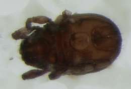
Figure 29.Diptacus berberinus sp. n.: A dorsal view of female B ventral view of female C lateral microtubercles D empodium E dorsal view of female posterior part F ventral view of female posterior part G leg I and leg II.
Vladimir Pešić, Ksenia A. Semenchenko, Wonchoel Lee
Zookeys
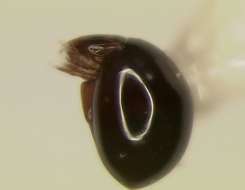
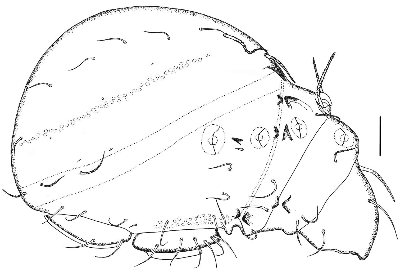
Figure 3.Torrenticola kimichungi sp. n., male holotype: A dorsal shield B ventral shield C ejaculatory complex D palp, lateral view E palp, medial view F gnathosoma. Scale bars = 100 μm.


