NMNH Plagiodinium belizeanum type specimen
Description :
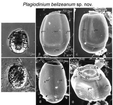
Figs 1-6. Plagiodinium belizeanum sp. nov. FIGS. 1-2. Light microscope view. Cells contain chloroplasts, spherical starch bodies (arrowheads) and a spherical posterior nucleus (n). FIG. 1. Cell is in left lateral view. FIG. 2. Cell is in right lateral view. FIGS.3-4. Cells viewed with the scanning electron microscope. FIG. 3. Cell is in left lateral view. FIG. 4. Cell is in right lateral view. Note an inclined, small, cap-shaped epitheca, deep cingulum, and an oblong hypotheca. The thecal surface is smooth. The large hypothecal plates 1'", 2'", 3'" (Fig. 3), and 4'" (Fig. 4) and antapical plate 1" are indicated. FIG. 5. The antapical 1" plate has a convex posterior shape. Small scattered thecal pores (arrowheads) are present. FIG. 6. Apical view of the epitheca. Epitheca narrowly oval pointed ventrally and rounded dorsally. The cingulum is deep, and the anterior and posterior margins are thickened. The cingulum has five plates (C1-C5) and is more depressed at its dorsal side. The hypothecal plates 1'", 2'", and 3'" are shown.
Inclus dans les pages suivantes :
- Life
- Cellular (Organismes cellulaires)
- Eukaryota (eucaryotes)
- SAR (Stramenopiles, Alveolates, Rhizaria)
- Alveolata
- Dinophyceae (Dinoflagellés)
- Prorocentrales
- Prorocentraceae
- Plagiodinium
- Plagiodinium belizeanum
- Dinoflagellata
Cette image ne figure dans aucune collection.
Informations sur la provenance
- licence
- cc-by-nc-sa-3.0
- droit d’auteur
- National Museum of Natural History, Smithsonian Institution
- original
- fichier de média d’origine
- site partenaire
- NMNH Marine Dinoflagellates
- ID