Image de Borrelia
Description :
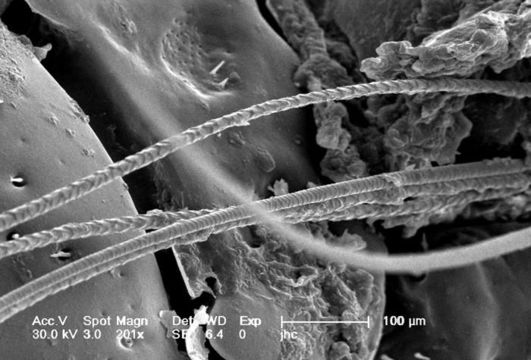
Under a magnification of 201X, this scanning electron micrographic (SEM) image depicted a dorsal view of an unidentified engorged female tick, which had been extracted from the skin of a pet cat while in the process of obtaining its blood meal. Note the presence of some of the cats fur, along with some of its skin tissue in which the ticks gnathosoma were still embedded. See PHIL 9972 and 9973 for additional, less magnified views of this scenario. It is from the basis capituli that the two spread pedipalps, and hidden skin-piercing hypostome and chelicerae emanate. On the dorsal surface of the basis capituli youll see two depressed areas known as the porose areas, through which secretions produced by dermal glands are released.
Created: 2006
Inclus dans les pages suivantes :
- Life
- Cellular (Organismes cellulaires)
- Bacteria
- Spirochaetes
- Spirochaetales
- Borrelia
- Borrelia burgdorferi
- Spirochaetaceae
- Spirochaetes
Cette image ne figure dans aucune collection.
Informations sur la provenance
- licence
- cc-publicdomain
- photographe
- Janice Carr
- fournisseur
- Public Health Image Library
- original
- fichier de média d’origine
- visiter la source
- site partenaire
- Public Health Image Library
- ID