Image de Pneumocystis
Description :
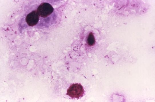
At a magnification of 1000X, this photomicrograph reveals Pneumocystis jirovecii fungi, which were present in this Giemsa-stained impression smear of rat lung tissue. Formerly known as Pneumocystis carinii, and classified as a protozoa, investigative tests upon this organisms nucleic acid and biochemical composition has since placed it in the Kingdom of Fungi.These fungi are found in the lungs of mammals where they reside without causing overt infection until the host's immune system becomes debilitated. Then, an oftentimes lethal pneumonia can result. Note the round cyst in the very middle of this image containing eight immature haploid neuclei, as well as numbers of freed trophozoites.
Created: 1977
Inclus dans les pages suivantes :
- Life
- Cellular
- Eukaryota
- Opisthokonta
- Nucletmycea
- Fungi
- Dikarya
- Ascomycota
- Pneumocystis
- Taphrinomycotina
- Pneumocystidomycetes
- Pneumocystidales
- Pneumocystidaceae
- Pneumocystis jirovecii
Cette image ne figure dans aucune collection.
Informations sur la provenance
- licence
- cc-publicdomain
- fournisseur
- Public Health Image Library
- original
- fichier de média d’origine
- visiter la source
- site partenaire
- Public Health Image Library
- ID