Image de Dasymutilla Ashmead 1899
Description :
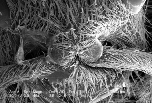
At a low magnification of only 46X, this scanning electron micrograph (SEM) showed the head region from an anterior view of a female velvet ant, Dasymutilla sp.. Note the two laterally positioned eyes which are partially visible at the topmost area of the photograph, but its the two anteriorly-placed antennae with their rounded "scapes" that are the most apparent head appendages. Like the antennae, the numerous hairs or setae adorning almost all of the insects exterior surfaces, act as sensory structures, supplying the organism with information about its environmental parameters. The jointed legs, from which the insects Phylum Arthropoda is derived, i.e., Arthro = jointed, and poda leg, are also partially visible, emanating from the thoracic region.
Created: 2007
Inclus dans les pages suivantes :
- Life
- Cellular (Organismes cellulaires)
- Eukaryota (eucaryotes)
- Opisthokonta
- Metazoa (animaux)
- Bilateria
- Protostomia
- Ecdysozoa
- Arthropoda (Arthropodes)
- Pancrustacea
- Hexapoda (Hexapodes)
- Insecta (Insectes)
- Pterygota (ptérygotes)
- Neoptera
- Endopterygota
- Hymenoptera
- Apocrita
- Aculeata
- Vespoidea
- Mutillidae
- Dasymutilla
- Panarthropoda
Cette image ne figure dans aucune collection.
Informations sur la provenance
- licence
- cc-publicdomain
- photographe
- Janice Carr
- fournisseur
- Public Health Image Library
- original
- fichier de média d’origine
- visiter la source
- site partenaire
- Public Health Image Library
- ID