Image de Orientia tsutsugamushi
Description :
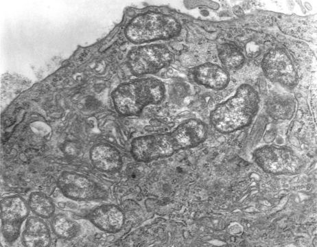
This 1976 transmission electron micrograph (TEM) depicted a hypertrophic peritoneal mesothelial cell of mouse that had been experimentally infected intraperitoneally with Orientia tsutsugamushi rickettsial micro-organisms. In this photomicrograph there were several organisms visible free within the mesothelial cell's cytoplasm.
Created: 1976
Inclus dans les pages suivantes :
- Life
- Cellular
- Bacteria
- Proteobacteria (Pseudomonadota)
- Alphaproteobacteria
- Rickettsiales
- Rickettsiaceae
- Orientia
- Orientia tsutsugamushi
Cette image ne figure dans aucune collection.
Informations sur la provenance
- licence
- cc-publicdomain
- fournisseur
- Public Health Image Library
- original
- fichier de média d’origine
- visiter la source
- site partenaire
- Public Health Image Library
- ID