""
Description :
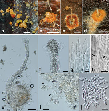
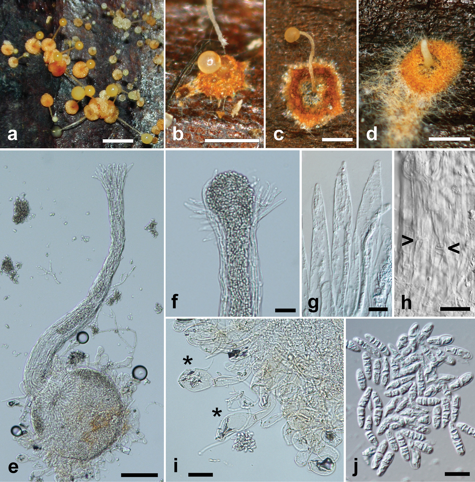
Figure 2.Pycnopulvinus aurantiacus (holotype PUL F2679). a Field photo of fresh sporocarps on palm leaf. Note the variable size and color of the sporocarps. Bar = 2 mm bâd Sporocarps of various stages and sizes after drying. Bars = 0.5 mm e Sporocarp as seen under the light microscope. Note the swollen pigmented base with surrounding large globose cells and the long tubular neck with a widening tip. Bar = 200 μm f Tip of the neck with spore mass exiting the sporocarp. Bar = 25 μm g Ostiolar hyphae at the tip of the neck. Bar = 10 μm h Outer surface of the neck, wide hyphae are visible, septa are marked with arrows. Bar = 10 μm i Globose cells (marked with asterisk) surrounding the base of the sporocarp. Bar = 25 μm j Multicellular spores produced inside the sporocarps. Note the four-celled spores on the left breaking into smaller compartments. Bar = 10 μm.
Inclus dans les pages suivantes :
- Life
- Cellular (Organismes cellulaires)
- Eukaryota (eucaryotes)
- Opisthokonta
- Nucletmycea
- Fungi
- Dikarya
- Basidiomycota
- Microbotryomycetes
- Heterogastridiales
- Heterogastridiaceae
- Pycnopulvinus
- Pycnopulvinus aurantiacus
Cette image ne figure dans aucune collection.
Informations sur la provenance
- licence
- cc-by-3.0
- droit d’auteur
- Merje Toome, M. Catherine Aime
- original
- fichier de média d’origine
- visiter la source
- site partenaire
- Mycokeys
- ID