Fig 1
Description :
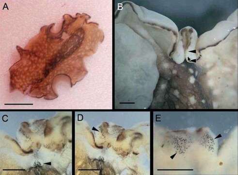
A, dorsal view of live specimen. B, detail of the anterior end. Arrow-heads show the interruption of the marginal black line at the apical region of the pseudotentacles. C, dorsal view of the anterior end with cerebral eyes (arrow-head). D, dorsal view of the anterior end; arrow head shows some of the ventral pseudotentacular eyes. E, ventral view of the anterior end with ventral pseudotentacular eyes (arrow heads). Scale bars: A: 5 mm; ?E: 1 mm.
Inclus dans les pages suivantes :
- Life
- Cellular (Organismes cellulaires)
- Eukaryota (eucaryotes)
- Opisthokonta
- Metazoa (animaux)
- Bilateria
- Protostomia
- Spiralia
- Platyhelminthes (Plathelminthes)
- unclassified Platyhelminthes
- Polycladida
- Pseudocerotoidea
- Pseudocerotidae
- Phrikoceros
- Phrikoceros mopsus
Cette image ne figure dans aucune collection.
Informations sur la provenance
- licence
- cc-by-nc-sa-4.0
- droit d’auteur
- WoRMS Editorial Board
- original
- fichier de média d’origine
- visiter la source
- site partenaire
- World Register of Marine Species
- ID