PHIL 3398 lores

Description :
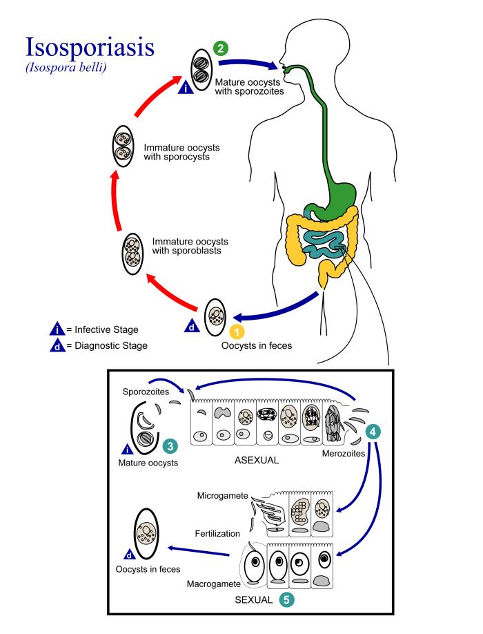
Summary.mw-parser-output table.commons-file-information-table,.mw-parser-output.fileinfotpl-type-information{border:1px solid #a2a9b1;background-color:#f8f9fa;padding:5px;font-size:95%;border-spacing:2px;box-sizing:border-box;margin:0;width:100%}.mw-parser-output table.commons-file-information-table>tbody>tr,.mw-parser-output.fileinfotpl-type-information>tbody>tr{vertical-align:top}.mw-parser-output table.commons-file-information-table>tbody>tr>td,.mw-parser-output table.commons-file-information-table>tbody>tr>th,.mw-parser-output.fileinfotpl-type-information>tbody>tr>td,.mw-parser-output.fileinfotpl-type-information>tbody>tr>th{padding:4px}.mw-parser-output.fileinfo-paramfield{background:#ccf;text-align:right;padding-right:0.4em;width:15%;font-weight:bold}.mw-parser-output.commons-file-information-table+table.commons-file-information-table,.mw-parser-output.commons-file-information-table+div.commons-file-information-table>table{border-top:0;padding-top:0;margin-top:-8px}@media only screen and (max-width:719px){.mw-parser-output table.commons-file-information-table,.mw-parser-output.commons-file-information-table.fileinfotpl-type-information{border-spacing:0;padding:0;word-break:break-word;width:100%!important}.mw-parser-output.commons-file-information-table>tbody,.mw-parser-output.fileinfotpl-type-information>tbody{display:block}.mw-parser-output.commons-file-information-table>tbody>tr>td,.mw-parser-output.commons-file-information-table>tbody>tr>th,.mw-parser-output.fileinfotpl-type-information>tbody>tr>td,.mw-parser-output.fileinfotpl-type-information>tbody>tr>th{padding:0.2em 0.4em;text-align:left;text-align:start}.mw-parser-output.commons-file-information-table>tbody>tr,.mw-parser-output.fileinfotpl-type-information>tbody>tr{display:flex;flex-direction:column}.mw-parser-output.commons-file-information-table+table.commons-file-information-table,.mw-parser-output.commons-file-information-table+div.commons-file-information-table>table{margin-top:-1px}.mw-parser-output.fileinfo-paramfield{box-sizing:border-box;flex:1 0 100%;width:100%}} Description: ID#:3398 This is an illustration of the life cycle of Isospora belli, the causal agent of Isosporiasis. At time of excretion, the immature oocyst contains usually one sporoblast (more rarely two) (1). In further maturation after excretion, the sporoblast divides in two (the oocyst now contains two sporoblasts); the sporoblasts secrete a cyst wall, thus becoming sporocysts; and the sporocysts divide twice to produce four sporozoites each (2). Infection occurs by ingestion of sporocysts-containing oocysts: the sporocysts excyst in the small intestine and release their sporozoites, which invade the epithelial cells and initiate schizogony (3). Upon rupture of the schizonts, the merozoites are released, invade new epithelial cells, and continue the cycle of asexual multiplication (4). Trophozoites develop into schizonts which contain multiple merozoites. After a minimum of one week, the sexual stage begins with the development of male and female gametocytes (5). Fertilization results in the development of oocysts that are excreted in the stool (1). Isospora belli infects both humans and animals. ([1]). Date: 2002. Source: CDC http://phil.cdc.gov/. Author: CDC/Alexander J. da Silva, PhD/Melanie Moser.
Inclus dans les pages suivantes :
- Life
- Cellular (Organismes cellulaires)
- Eukaryota (eucaryotes)
- SAR (Stramenopiles, Alveolates, Rhizaria)
- Alveolata
- Apicomplexa
- Conoidasida
- Coccidia (Coccidie)
- Eucoccidiorida
- Eimeriorina
- Sarcocystidae
- Cystoisospora
- Cystoisospora belli
Cette image ne figure dans aucune collection.
Informations sur la provenance
- licence
- cc-publicdomain
- créateur
- CDC/Alexander J. da Silva, PhD/Melanie Moser
- source
- CDC http://phil.cdc.gov/
- original
- fichier de média d’origine
- visiter la source
- site partenaire
- Wikimedia Commons
- ID