Image de Dicondylia
Description :
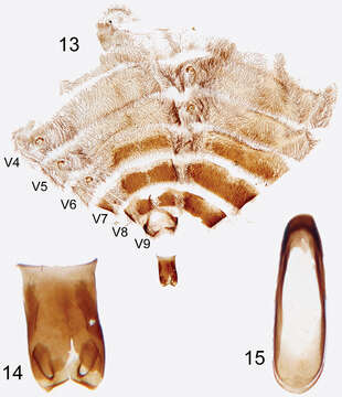
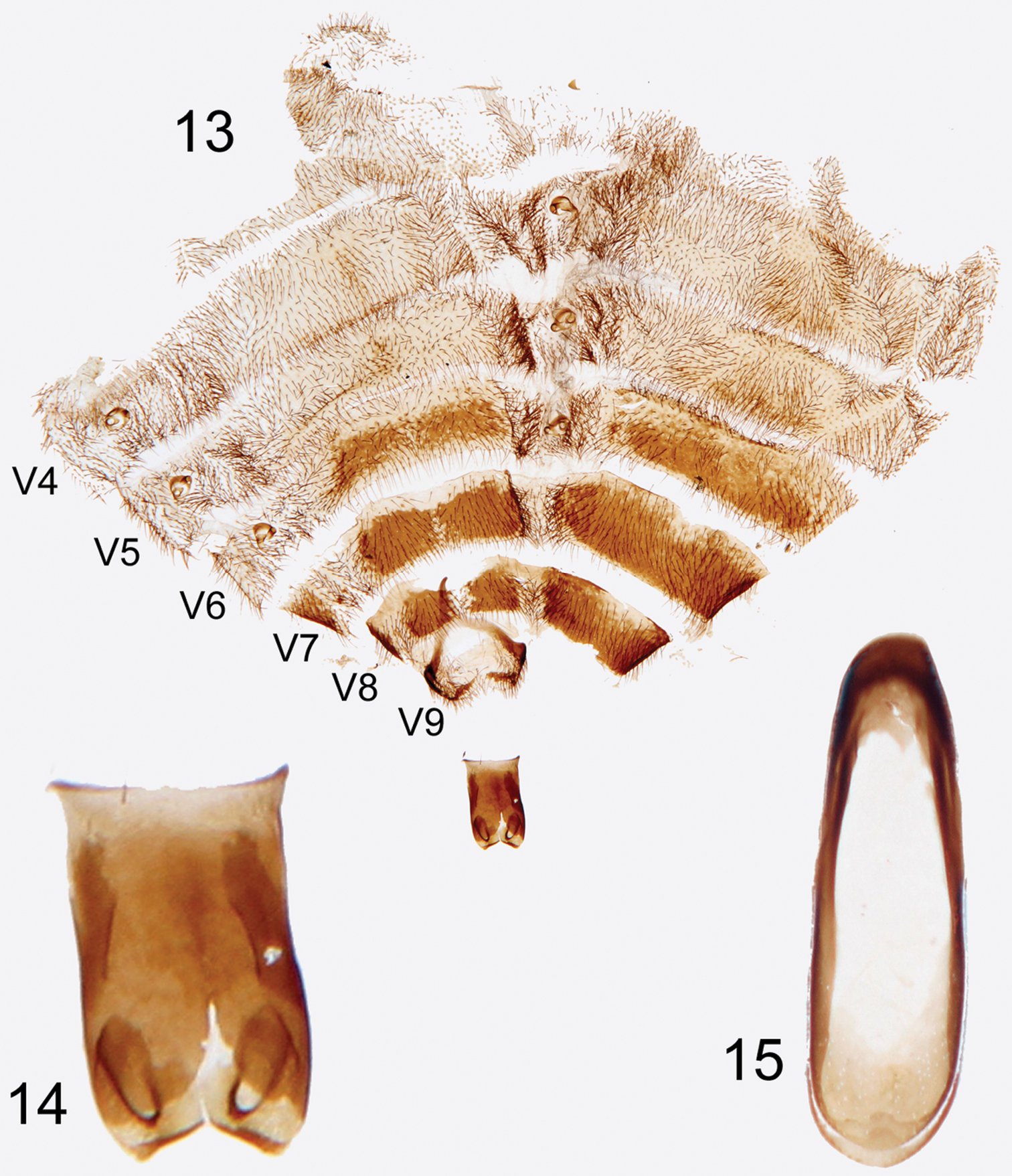
Figures 13–15.Photographs of slide mounted abdominal structures from holotype male of Rhipidocyrtus muiri Falin & Engel, gen. et sp. n. 13 Splayed abdomen as preserved on slide, numbered ventrites to the left, unnumbered tergites to the right 14 Enlarged detail, ventral view of tegmen 15 Enlarged detail of median lobe.
Inclus dans les pages suivantes :
- Biota
- Eukaryota (eucaryotes)
- Unikonta
- Opisthokonta
- Metazoa (animaux)
- Epitheliozoa
- Eumetazoa
- Bilateria
- Protostomia
- Ecdysozoa
- Arthropoda (Arthropodes)
- Hexapoda (Hexapodes)
- Insecta (Insectes)
- Dicondylia
- Pterygota (ptérygotes)
- Metapterygota
- Neoptera
- Eumetabola
- Endopterygota
- Coleoptera (Coléoptère)
- Polyphaga
- Cucujiformia
- Tenebrionoidea
- Ripiphoridae
- Ripidiinae
- Ripidiini
- Rhipidocyrtus
- Rhipidocyrtus muiri
Cette image ne figure dans aucune collection.
Informations sur la provenance
- licence
- cc-by-3.0
- droit d’auteur
- Zachary H. Falin, Michael S. Engel
- citation bibliographique
- Falin Z, Engel M (2014) Serendipity at the Smithsonian: The 107-year journey of Rhipidocyrtus muiri Falin & Engel, new genus and species (Ripidiinae, Ripidiini), from jungle beast to valid taxon ZooKeys 424: 101–116
- original
- fichier de média d’origine
- visiter la source
- site partenaire
- Zookeys
- ID