Image de Xyphinus
Description :
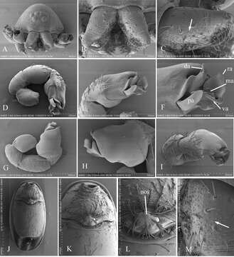
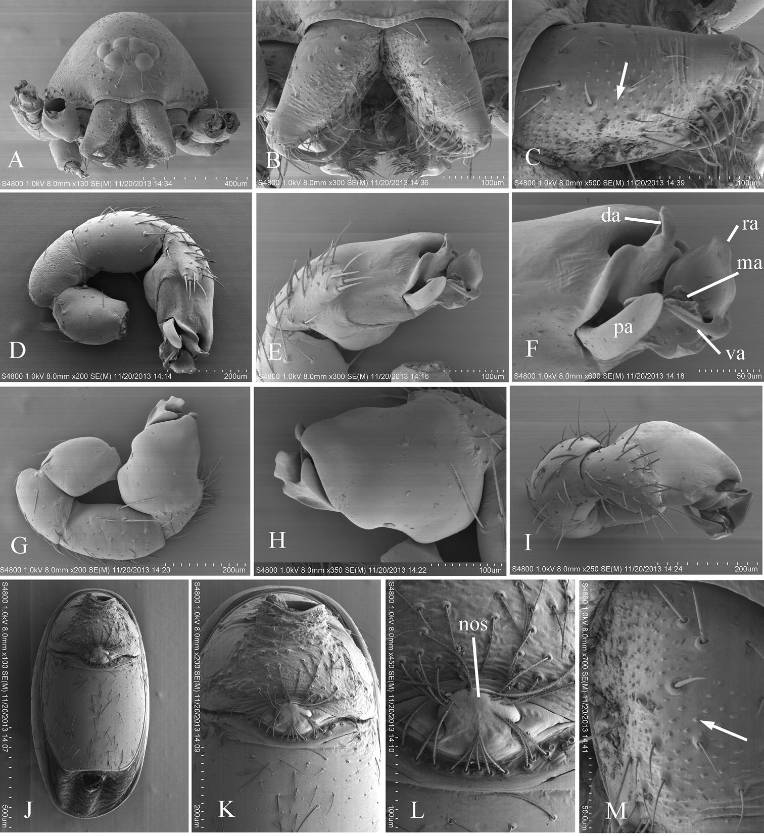
Figure 6.Xyphinus hwangi sp. n., SEM. A male prosoma, anterior view B, C, M male chelicerae, frontal view (arrow shows the small granules) D, G, I male left palp, prolateral, retrolateral and dorsal views E, H male left palpal bulb, prolateral and retrolateral views F distal part of male left palpal bulb, prolateral view J female abdomen, ventral view K, L female genital area, ventral view. Abbreviations: da = dorsal apophysis; ma = medial apophysis; nos = nose-shaped protuberance; pa = prolateral apophysis; ra = retrolateral apophysis; va = ventral apophysis.
Inclus dans les pages suivantes :
- Life
- Cellular (Organismes cellulaires)
- Eukaryota (eucaryotes)
- Opisthokonta
- Metazoa (animaux)
- Bilateria
- Protostomia
- Ecdysozoa
- Arthropoda (Arthropodes)
- Chelicerata (Chélicérates)
- Arachnida (Arachnide)
- Araneae (Aranéides)
- Opisthothelae
- Araneomorphae (Aranéomorphes)
- Haplogynae
- Oonopidae
- Xyphinus
- Xyphinus hwangi
- Panarthropoda
Cette image ne figure dans aucune collection.
Informations sur la provenance
- licence
- cc-by-3.0
- droit d’auteur
- Yanfeng Tong, Shuqiang Li
- citation bibliographique
- Tong Y, Li S (2014) A survey of oonopid spiders in Taiwan with descriptions of three new species ZooKeys 396: 67–86
- original
- fichier de média d’origine
- visiter la source
- site partenaire
- Zookeys
- ID