Image de Scalidophora

Description :
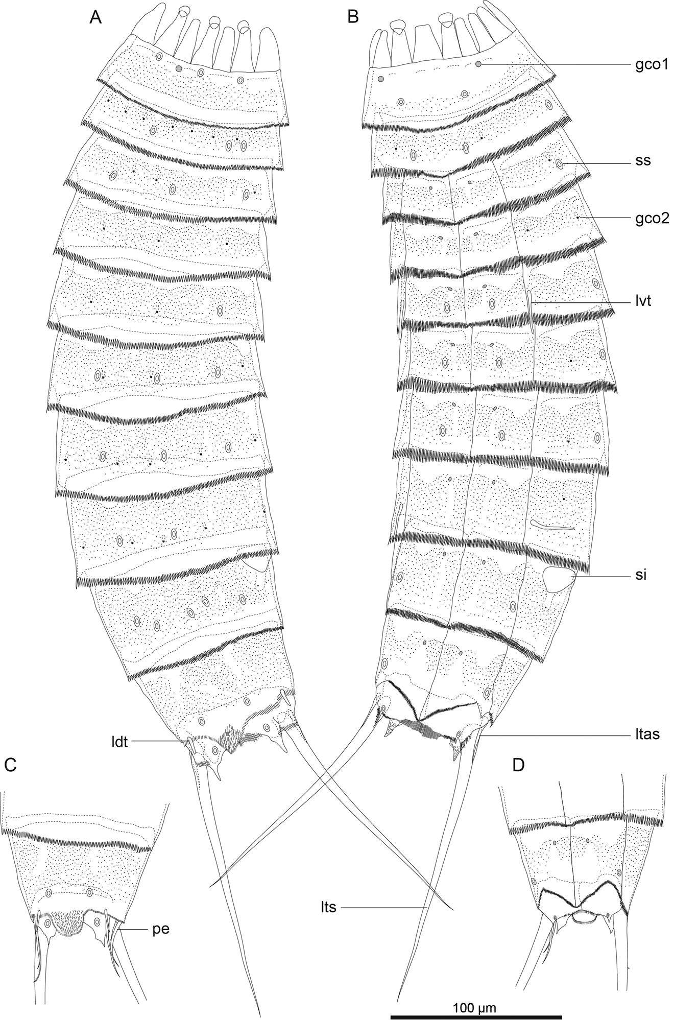
Figure 2.Echinoderes komatsui sp. n., camera lucida drawings. A, B Holotype, female (RUMF-ZK-00001), entire animal, dorsal and ventral views, respectively C, D allotype, male (RUMF-ZK-00002), segments 9–11, dorsal and ventral views, respectively. Double circle, grey circle, and black circle indicate sensory spot, type 1 glandular cell outlet, and type 2 glandular cell outlet, respectively. Abbreviations: gco1, type 1 glandular cell outlet; gco2, type 2 glandular cell outlet; ldt, laterodorsal tubule; ltas, lateral terminal accessory spine; lts, lateral terminal spine; lvt, lateroventral tubule; pe, penile spine; si, sieve plate; ss, sensory spot.
Inclus dans les pages suivantes :
- Eukaryota (eucaryotes)
- Opisthokonta
- Metazoa (animaux)
- Bilateria
- Protostomia
- Ecdysozoa
- Scalidophora
- Life
- Cellular (Organismes cellulaires)
- Kinorhyncha (Kinorhynques)
- Cyclorhagida
- Echinoderidae
- Echinoderes
- Echinoderes komatsui
Cette image ne figure dans aucune collection.
Informations sur la provenance
- licence
- cc-by-3.0
- droit d’auteur
- Hiroshi Yamasaki, Shinta Fujimoto
- citation bibliographique
- Yamasaki H, Fujimoto S (2014) Two new species in the Echinoderes coulli group (Echinoderidae, Cyclorhagida, Kinorhyncha) from the Ryukyu Islands, Japan ZooKeys 382: 27–52
- original
- fichier de média d’origine
- visiter la source
- site partenaire
- Zookeys
- ID