Thallus detail, Multi layer image (DOF)
Description :
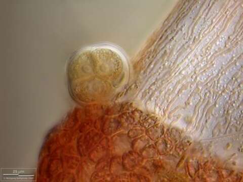
This detail view of a Ceramium diaphanum thallus segment shows the two cell types of the thallus: one tall axial cell (light, with tubular rhodoplasts) and many little reddish cortical cells with their lenticular rhodoplasts. In the tetrasporangium three cells with their nuclei are visible.
Collected from Bodden, the brackish waters lying between the isles of Hiddensee and Ruegen (German Baltic Sea). This image was taken using Zeiss Universal with Olympus C7070 CCD camera.
Inclus dans les pages suivantes :
- Eukaryota (eucaryotes)
- Eurhodophytina
- Life
- Cellular (Organismes cellulaires)
- Archaeplastida
- Rhodophyta
- Florideophyceae
- Rhodymeniophycidae
- Ceramiales
- Ceramiaceae
- Ceramium
- Ceramium diaphanum
Cette image ne figure dans aucune collection.
Informations sur la provenance
- licence
- cc-by-nc
- fournisseur
- micro*scope
- original
- fichier de média d’origine
- visiter la source
- site partenaire
- micro*scope
- ID