in vivo;left side
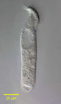
Description :
Left lateral view of Metopus palaeformis (Kahl, 1927).Synonyms probably include Tesnospira alba (Jankowski,1964),M. hyalinus (Kahl,19270 and M. tenuis (Kahl,1927) among others.Morphology is highly variable probably explaining the large number of synonyms. The cell is flask-shaped to elongate (as in this example).The anterior end is twisted to the left resulting in a rounded lip that overhangs the peristome.The spiral peristome is bordered on the left by an adoral zone of membranelles and on the right by five closely spaced kineties,the "perizonal stripe".Just to the right of the posterior termination of the AZM is a short, inconspicuous undulating membrane(usually visible only in silver-stained preparations).The The right somatic kineties parallel the peristome anteriorly and the left somatic kineties terminate at the margin of the peristome.There is no long tuft of caudal cilia. The prominent ellipsoid macronucleus and adjacent micronucleus are in the anterior half. The contractile vacuole is at the posterior end.The cytoplasm contains endosymbiotic methanogenic bacilli (not seen here).There is an aggregate of brown refractile granules at the anterior end typical of the metopid ciliates.Collected from the bottom sediments of an organically enriched rain pool with abundant decaying grass contaminated by Canada goose (Branta canadensis) droppings.Boise, Idaho. January 2006. DIC.
Inclus dans les pages suivantes :
- Life
- Cellular (Organismes cellulaires)
- Eukaryota (eucaryotes)
- SAR (Stramenopiles, Alveolates, Rhizaria)
- Alveolata
- Ciliophora
- Intramacronucleata
- Armophorea
- Metopida
- Metopidae
- Metopus
- Metopus palaeformis
Cette image ne figure dans aucune collection.
Informations sur la provenance
- licence
- cc-by-nc
- auteur
- William Bourland
- fournisseur
- micro*scope
- original
- fichier de média d’origine
- visiter la source
- site partenaire
- micro*scope
- ID