in vivo; right ventrolateral view
Description :
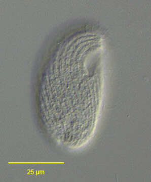
Right ventrolateral view of the colpodid ciliate, Bryometopus atypicus (Foissner,1980). Similar in overall shape to Colpoda maupasi. The dorsum of the cell is convex and the ventral surface straight. The subapical cytostome occupies the anterior 1/4 of the cell length.It is slightly oblique to the long axis of the cell. The somatic kineties (composed of dikinetids) are moderately spiralled curving around the cytostome to end on a short preoral suture.Approximately 7 postoral kineties terminate on the left border of the cytostome.There is a slightly curved right paraoral membrane and an adoral zone of membranelles on the left border of the cytostome.Rows of mucocysts occur between somatic kineties.The spherical macronucleus and adjacent single micronucleus is in the cell center.The posterior contractile vacuole has a distinctive large cylindrical excretory pore.Zoochlorellae are absent.Collected from an organically enriched rainwater pool with abundant decaying grass in Boise, Idaho. January 2006.DIC.
Inclus dans les pages suivantes :
- Life
- Cellular (Organismes cellulaires)
- Eukaryota (eucaryotes)
- SAR (Stramenopiles, Alveolates, Rhizaria)
- Alveolata
- Ciliophora
- Intramacronucleata
- Colpodea
- Bryometopida
- Bryometopidae
- Bryometopus
- Bryometopus atypicus
Cette image ne figure dans aucune collection.
Informations sur la provenance
- licence
- cc-by-nc
- auteur
- William Bourland
- fournisseur
- micro*scope
- original
- fichier de média d’origine
- visiter la source
- site partenaire
- micro*scope
- ID