Ventral infraciliature, late division
Description :
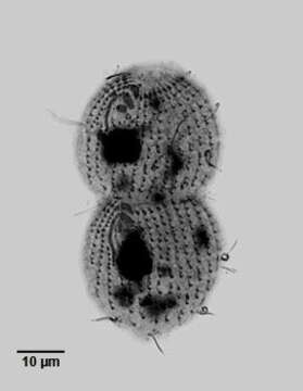
Ventral view of the infraciliature of late division of the hymenostome ciliate, Dexiotricha granulosa (Kent, 1881) Foissner, 1994. Synonym of Loxocephulus granulosa. The cell is ovoid, broadly rounded posteriorly and truncate anteriorly. Regular longitudinal kineties terminate at a subapical band of circumferential kineties demarcating a cilia-free truncate apical area or frontal plate. Fibrils radiate anteriorly from the kinetids of the anteriormost paratene (seen here). There is a single long caudal cilium. The oral aperture is small and difficult to visualize in vivo. It is located in the anterior quarter with an undulating membrane on the right (seen here) and 3 membranelles ( seen most clearly in the proter). The macronucleus is spheroid and located in the mid-cell. Single contractile vacuole. From freshwater pond near Boise, Idaho. Silver carbonate stain (see Foissner, W. Europ. J. Protistol., 27:313-330;1991).Brightfield. Black and white.
Inclus dans les pages suivantes :
- Life
- Cellular (Organismes cellulaires)
- Eukaryota (eucaryotes)
- SAR (Stramenopiles, Alveolates, Rhizaria)
- Alveolata
- Ciliophora
- Intramacronucleata
- Oligohymenophorea
- Scuticociliatia
- Philasterida
- Loxocephalidae
- Dexiotricha
- Dexiotricha granulosa
Cette image ne figure dans aucune collection.
Informations sur la provenance
- licence
- cc-by-nc
- auteur
- William Bourland
- fournisseur
- micro*scope
- original
- fichier de média d’origine
- visiter la source
- site partenaire
- micro*scope
- ID