cytopharyngeal folds
Description :
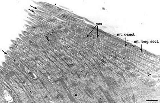
A view of the cytopharynx but in a dividing Didinium. This cytopharynx is in the proter (anterior) daughter cell. The lamellae, each consisting of a set of perpendicularly arranged microtubules (mt), cover this food-vacuole forming region. Vesicles (ves) lie near the cytopharyngeal membrane and fuse with the membrane (arrows). EM taken on 5/20/69 by R. Allen with Philips 300 TEM. Neg. 6,370X. Bar = 1 micron.This image is available in Richard Allen's collection.
Inclus dans les pages suivantes :
- Life
- Cellular (Organismes cellulaires)
- Eukaryota (eucaryotes)
- SAR (Stramenopiles, Alveolates, Rhizaria)
- Alveolata
- Ciliophora
- Intramacronucleata
- Litostomatea
- Haptoria
- Haptorida
- Didiniidae
- Didinium
- Didinium nasutum
Cette image ne figure dans aucune collection.
Informations sur la provenance
- licence
- cc-by-nc
- auteur
- R. D. Allen
- fournisseur
- micro*scope
- original
- fichier de média d’origine
- visiter la source
- site partenaire
- micro*scope
- ID