encystment
Description :
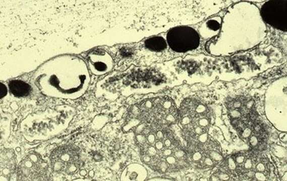
Transmission electron micrograph of a thin section through the surface of an encysting cell. The encystment process has just started - as can be seen from the sintered appearance of the siliceous scales. There is a layer of mucus outside the cell, extrusomes under the cell surface, and many mitochondria below the siliceous scales.
Inclus dans les pages suivantes :
- Life
- Cellular (Organismes cellulaires)
- Eukaryota (eucaryotes)
- SAR (Stramenopiles, Alveolates, Rhizaria)
- Stramenopiles (Heterokonta)
- Oomycota
- Actinophryida
- Actinophrys
- Actinophrys sol
Cette image ne figure dans aucune collection.
Informations sur la provenance
- licence
- cc-by-nc
- auteur
- D. J. Patterson.
- fournisseur
- micro*scope
- original
- fichier de média d’origine
- visiter la source
- site partenaire
- micro*scope
- ID