in vivo;ventral surface
Description :
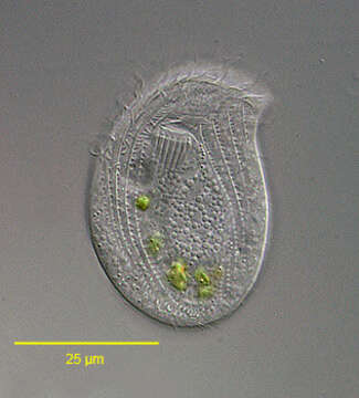
Portrait (ventral surface) of the chilodonellid ciliate Pseudochilodonopsis piscatoris (Blochmann, 1895) Foissner, 1979. The cell is drawn out to the left in a distinct pointed preoral beak. The posterior is broadly rounded. The cell is strongly dorsoventrally compressed. The dorsum is slightly domed and the ventral surface flat. The ciliature is reduced to the ventral surface except for a distinctive dorsal brush which is set back from the anterior edge of the cell and arches across the nearly its entire width. The ventral ciliature consists of right (5) and left (6) kineties separated by a wide bare postoral area. There are two circumoral kineties and a fragmented preoral kinety. The anterior ends of the left somatic kineties abut the transversely oriented fragments of the preoral kinety. These fragments ascend stair-step fashion to the tip of the beak. The cytostome, situated in the anterior 1/4 of the cell, is supported by nematodesmata forming a cyrtos. There are two contractile vacuoles. The nucleus is heteromerous. The genus Pseudochilodonopsis is distinguished from members of the similar genus, Chilodonella, by the fragmented preoral kinety and the long arched dorsal brush. Both features are difficult to appreciate without DIC optics or silver impregnation techniques. P. piscatoris feeds on green algae and diatoms. It is usually found in the surface film of samples collected from a freshwater pond near Boise,Idaho. February 2005. DIC.
Inclus dans les pages suivantes :
- Life
- Cellular (Organismes cellulaires)
- Eukaryota (eucaryotes)
- SAR (Stramenopiles, Alveolates, Rhizaria)
- Alveolata
- Ciliophora
- Intramacronucleata
- Phyllopharyngea
- Phyllopharyngia
- Chlamydodontida
- Chilodonellidae
- Pseudochilodonopsis
- Pseudochilodonopsis piscatoris
Cette image ne figure dans aucune collection.
Informations sur la provenance
- licence
- cc-by-nc
- auteur
- William Bourland
- fournisseur
- micro*scope
- original
- fichier de média d’origine
- visiter la source
- site partenaire
- micro*scope
- ID