oral region
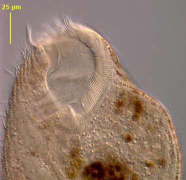
Description :
Anterior detail of the marine heterotrich ciliate, Condylostoma arenarium (Spiegel, 1926). The dorsoventrally flattened elongate cell body is very contractile (this cell is partly contracted). Contraction is probably mediated by calcium dependent subcortical myonemes and cell extension by interaction of cortical postciliary microtubular ribbons. The broad anterior V-shaped peristome is bordered on the right by a large undulating membrane. An adoral zone of membranelles (AZM) winds from right anterior clockwise around the left margin of the peristome. Several cirri are located at the right-most end of the AZM (seen well here). Uniform longitudinal somatic kineties are separated by strips of yellowish cortical granules (pigmentocysts). Pigmentocysts are extrusomes containing toxic substances. They play a role in cell defense against predators and may also function in photoreception. Pigmentocysts are also found in other heterotrichs (e.g. Blepharisma and Stentor species). The long moniliform macronucleus extends along the right cell margin (faintly visible here). No contractile vacuole. Brownish food vacuoles throughout the cytoplasm in this individual contain ingested dinoflagellates (Amphidinium). Collected from a seawater aquarium in Boise, Idaho January 2004. DIC optics.
Inclus dans les pages suivantes :
- Life
- Cellular (Organismes cellulaires)
- Eukaryota (eucaryotes)
- SAR (Stramenopiles, Alveolates, Rhizaria)
- Alveolata
- Ciliophora
- Postciliodesmatophora
- Heterotrichea
- Heterotrichida
- Condylostomatidae
- Predurostyla
- Predurostyla arenaria
Cette image ne figure dans aucune collection.
Informations sur la provenance
- licence
- cc-by-nc
- auteur
- Bill Bourland
- fournisseur
- micro*scope
- original
- fichier de média d’origine
- visiter la source
- site partenaire
- micro*scope
- ID