-
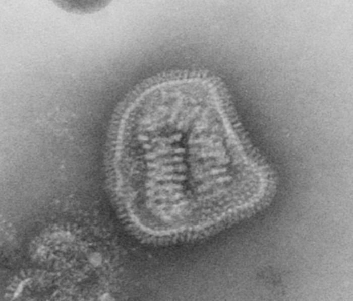
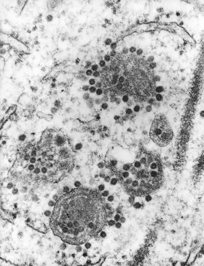
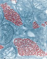
This negative-stained transmission electron micrograph (TEM) depicts the ultrastructural details of an influenza virus particle, or virion. A member of the taxonomic family Orthomyxoviridae, the influenza virus is a single-stranded RNA organismThe flu is a contagious respiratory illness caused by influenza viruses. It can cause mild to severe illness, and at times can lead to death. The best way to prevent this illness is by getting a flu vaccination each fall.Every year in the United States, on average:- 5% to 20% of the population gets the flu- more than 200,000 people are hospitalized from flu complications, and- about 36,000 people die from flu. Some people, such as older people, young children, and people with certain health conditions, are at high risk for serious flu complications. See PHIL 10073 for a colorized version of this image.Created: 1981
-
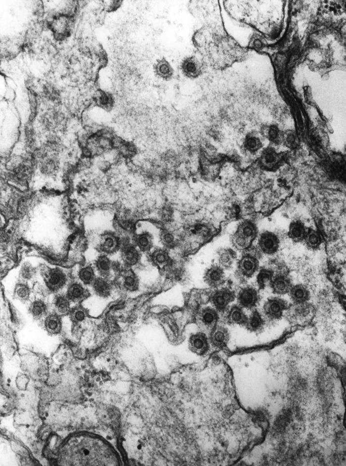
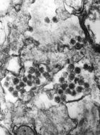
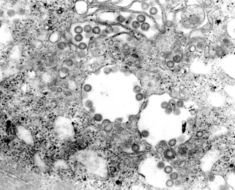
This negatively-stained transmission electron micrograph (TEM) revealed the presence of numerous doulble-stranded, full-length DNA-containing (dsDNA) spumavirus, or foamyvirus virions. One of the identifying morphologic characteristics displayed by these virions is the spikey nature of their protective proteinaceous capsid, which is a feature evident in this TEM.Created: 1975
-
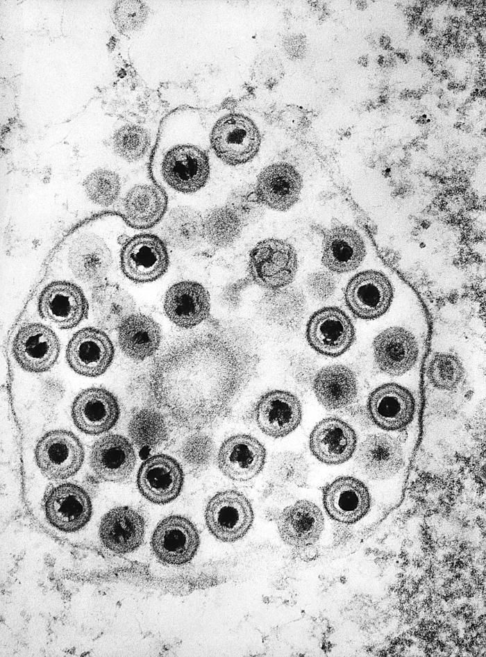
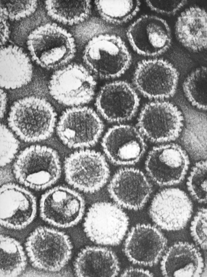
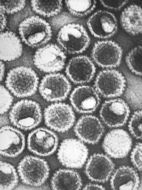
This negatively-stained transmission electron micrograph (TEM) revealed the presence of numerous herpes simplex virions, members of the Herpesviridae virus family. There are two strains of the herpes simplex virus, HSV-1, which is responsible for cold sores, and HSV-2, which is responsible for genital herpes. At the core of its icosahedral proteinaceous capsid, the HSV contains a double-stranded DNA linear genome.Created: 1975
-
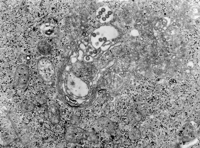
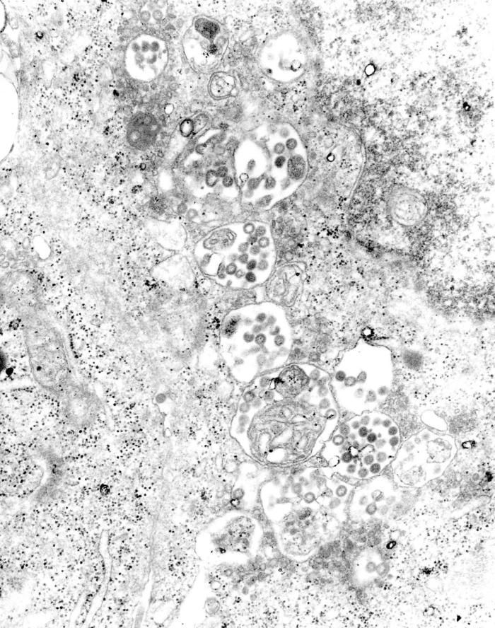
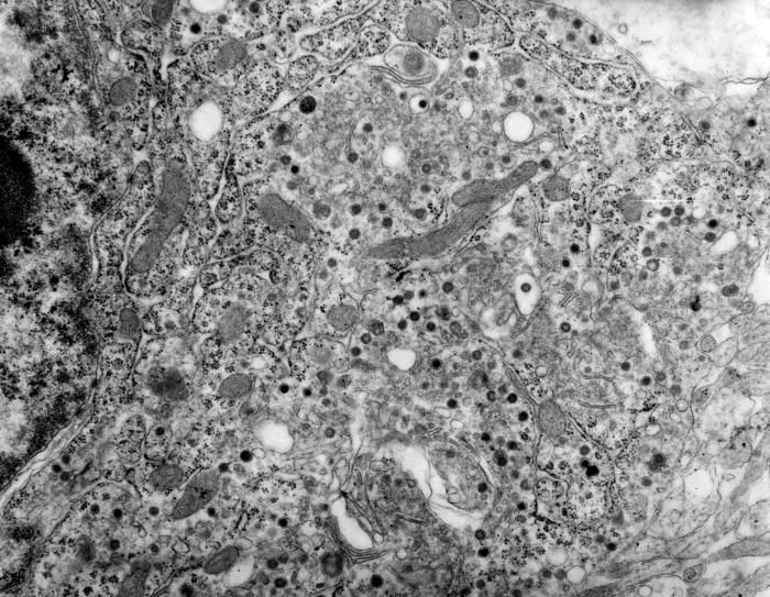
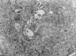
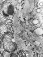
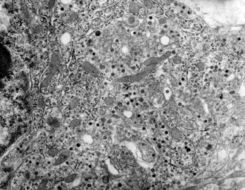
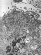
This transmission electron micrograph (TEM) depicted a highly magnified view of a tissue that had been infected with Rift Valley fever (RVF) virus. RVF virus is a member of the genus Phlebovirus in the family Bunyaviridae and was first reported in livestock in Kenya around 1900. It is found to be an acute, fever-causing viral disease that affects domestic animals (such as cattle, buffalo, sheep, goats, and camels) and humans. RVF is most commonly associated with mosquito-borne epidemics during years of unusually heavy rainfall.Created:
-
This negatively-stained transmission electron micrograph (TEM) revealed the presence of numerous doulble-stranded, full-length DNA-containing (dsDNA) spumavirus, or foamyvirus virions. One of the identifying morphologic characteristics displayed by these virions is the spikey nature of their protective proteinaceous capsid, which is a feature evident in this TEM.Created: 1975
-
This negatively-stained transmission electron micrograph (TEM) revealed the presence of numerous herpes simplex virions, members of the Herpesviridae virus family. There are two strains of the herpes simplex virus, HSV-1, which is responsible for cold sores, and HSV-2, which is responsible for genital herpes. At the core of its icosahedral proteinaceous capsid, the HSV contains a double-stranded DNA linear genome.Created: 1975
-
This transmission electron micrograph (TEM) depicted a highly magnified view of a tissue that had been infected with Rift Valley fever (RVF) virus. RVF virus is a member of the genus Phlebovirus in the family Bunyaviridae and was first reported in livestock in Kenya around 1900. It is found to be an acute, fever-causing viral disease that affects domestic animals (such as cattle, buffalo, sheep, goats, and camels) and humans. RVF is most commonly associated with mosquito-borne epidemics during years of unusually heavy rainfall.Created:
-
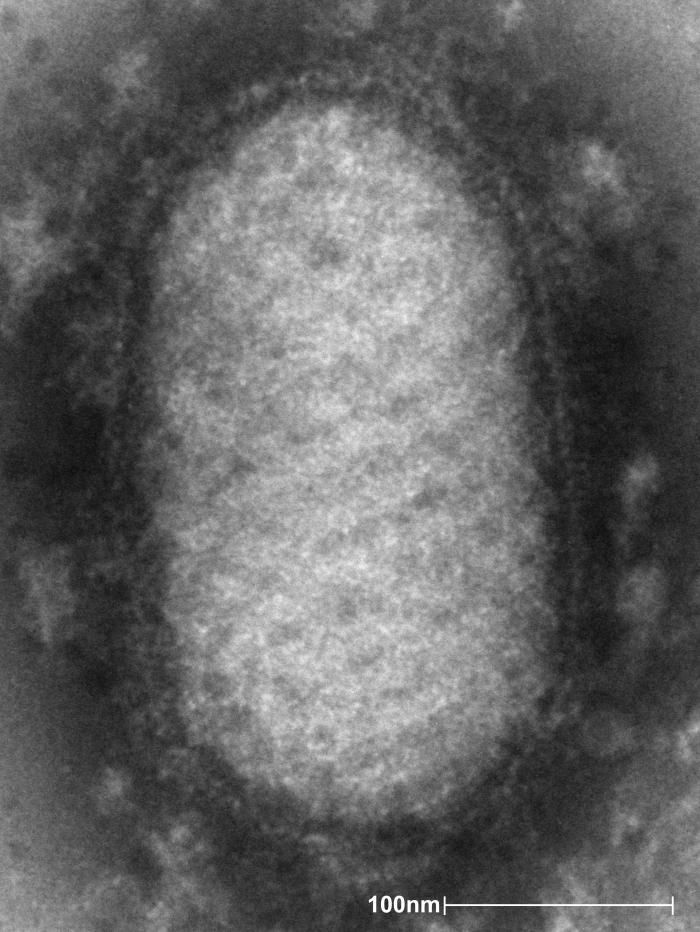
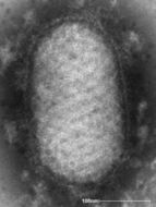
This negative-stained transmission electron micrograph (TEM) image depicted the ultrastructural details of an Orf virus, a member of the genus Parapoxvirus. Note the spiral arrangement of the external tubular ridges on the ovoid-shaped virus particle, or virion.Created:
-
This transmission electron micrograph (TEM) depicted a highly magnified view of a tissue that had been infected with Rift Valley fever (RVF) virus. RVF virus is a member of the genus Phlebovirus in the family Bunyaviridae and was first reported in livestock in Kenya around 1900. It is found to be an acute, fever-causing viral disease that affects domestic animals (such as cattle, buffalo, sheep, goats, and camels) and humans. RVF is most commonly associated with mosquito-borne epidemics during years of unusually heavy rainfall.Created:
-
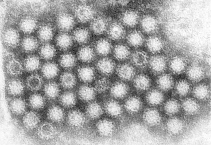
This transmission electron micrograph (TEM) revealed some of the ultrastructural morphology displayed by norovirus virions, or virus particles.Noroviruses belong to the genus Norovirus, and the family Caliciviridae. They are a group of related, single-stranded RNA, nonenveloped viruses that cause acute gastroenteritis in humans. Norovirus was recently approved as the official genus name for the group of viruses provisionally described as Norwalk-like viruses (NLV). See PHIL 10708 for a colorized version of this image.Created:
-
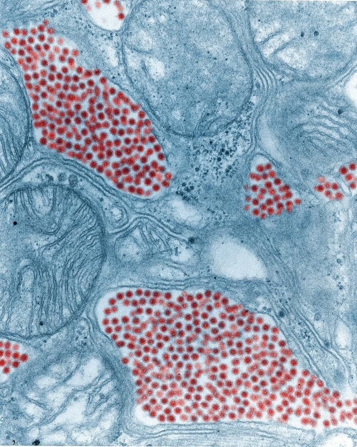
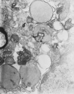
Magnified approximately 34,000x, this transmission electron micrograph (TEM) depicted a tissue section that had been infected with Rift Valley fever (RVF) virus. RVF virus is a member of the genus Phlebovirus in the family Bunyaviridae and was first reported in livestock in Kenya around 1900. It is found to be an acute, fever-causing viral disease that affects domestic animals (such as cattle, buffalo, sheep, goats, and camels) and humans. RVF is most commonly associated with mosquito-borne epidemics during years of unusually heavy rainfall.Created:
-
This transmission electron micrograph (TEM) revealed some of the ultrastructural morphology displayed by norovirus virions, or virus particles.Noroviruses belong to the genus Norovirus, and the family Caliciviridae. They are a group of related, single-stranded RNA, nonenveloped viruses that cause acute gastroenteritis in humans. Norovirus was recently approved as the official genus name for the group of viruses provisionally described as Norwalk-like viruses (NLV). See PHIL 10706 for a colorized version of this image.Created:
-
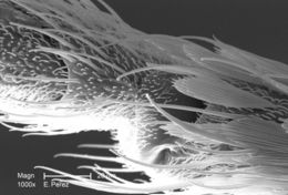
At a magnification of 1000X, twice that of PHIL 10557, this scanning electron micrograph (SEM) revealed some of the minute exoskeletal details found at the proboscis tip of an unidentified mosquito found deceased in the suburbs of Decatur, Georgia. The proboscis is the organ used by this, as well as other like insects, to feed upon the blood of a warm-blooded host, including human beings. What you see here, is the sheath that encases a pair of needle-sharp "stylets", which together are known as the "fascicle". The larger of the two stylets, known as the "labrum", when viewed in cross-section, takes on the shape of a "V", and acts as a gutter, which directs the ingested host blood towards the insect's mouth. The hair-like structures are known as "setae", and are really extensions of the insect's exoskeletal, chitinous covering. These setae act as sensory organs, transmitting impulses indicating changes in the organism's environment.Created: 2008
-
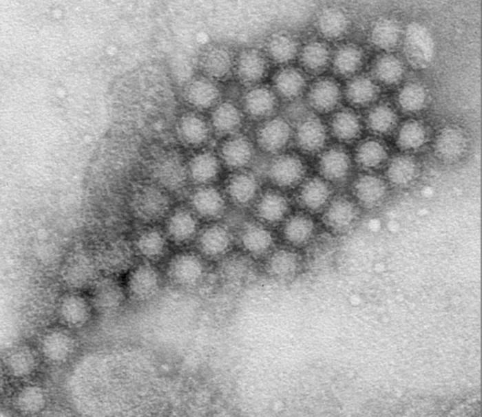
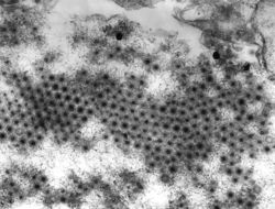
This transmission electron micrograph (TEM) revealed the presence of numbers of Nodamura virus virions. At its core, the Nodamura virus possesses a positive-sense, single-stranded RNA virus ((+) ssRNA) genome. Its outer protein coat, or capsid, is icosahedral (T=3) in shape, which means that its composed of 32 equilateral, triangular facets.Created: 1975
-
At a magnification of 1000X, twice that of PHIL 10557, this scanning electron micrograph (SEM) revealed some of the minute exoskeletal details found at the proboscis tip of an unidentified mosquito found deceased in the suburbs of Decatur, Georgia. The proboscis is the organ used by this, as well as other like insects, to feed upon the blood of a warm-blooded host, including human beings. What you see here, is the sheath that encases a pair of needle-sharp "stylets", which together are known as the "fascicle". The larger of the two stylets, known as the "labrum", when viewed in cross-section, takes on the shape of a "V", and acts as a gutter, which directs the ingested host blood towards the insect's mouth. The hair-like structures are known as "setae", and are really extensions of the insect's exoskeletal, chitinous covering. These setae act as sensory organs, transmitting impulses indicating changes in the organism's environment.Created: 2008
-
This transmission electron micrograph (TEM) revealed the presence of numbers of Nodamura virus virions. At its core, the Nodamura virus possesses a positive-sense, single-stranded RNA virus ((+) ssRNA) genome. Its outer protein coat, or capsid, is icosahedral (T=3) in shape, which means that its composed of 32 equilateral, triangular facets.Created: 1975
-
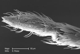
Magnified 500X, this scanning electron micrograph (SEM) revealed some of the minute exoskeletal details found at the proboscis tip of an unidentified mosquito found deceased in the suburbs of Decatur, Georgia. The proboscis is the organ used by this, as well as other like insects, to feed upon the blood of a warm-blooded host, including human beings. What you see here, is the sheath that encases a pair of needle-sharp "stylets", which together are known as the "fascicle". The larger of the two stylets, known as the "labrum", when viewed in cross-section, takes on the shape of a "V", and acts as a gutter, which directs the ingested host blood towards the insect's mouth. The hair-like structures are known as "setae", and are really extensions of the insect's exoskeletal, chitinous covering. These setae act as sensory organs, transmitting impulses indicating changes in the organism's environment.Created: 2008
-
This transmission electron micrograph (TEM) revealed the presence of numbers of Nodamura virus virions. At its core, the Nodamura virus possesses a positive-sense, single-stranded RNA virus ((+) ssRNA) genome. Its outer protein coat, or capsid, is icosahedral (T=3) in shape, which means that its composed of 32 equilateral, triangular facets.Created: 1975
-
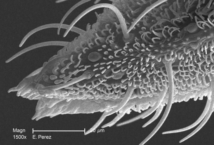
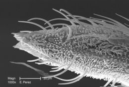
Magnified 1500X, this scanning electron micrograph (SEM) revealed some of the minute exoskeletal details found at the proboscis tip of an unidentified mosquito found deceased in the suburbs of Decatur, Georgia. The proboscis is the organ used by this, as well as other like insects, to feed upon the blood of a warm-blooded host, including human beings. What you see here, is the sheath that encases a pair of needle-sharp "stylets", which together are known as the "fascicle". The larger of the two stylets, known as the "labrum", when viewed in cross-section, takes on the shape of a "V", and acts as a gutter, directing the ingested host blood towards the insect's mouth. The hair-like structures are known as "setae", and are really extensions of the insect's exoskeletal, chitinous covering. These setae act as sensory organs, transmitting impulses indicating changes in the organism's environment.Created: 2008
-
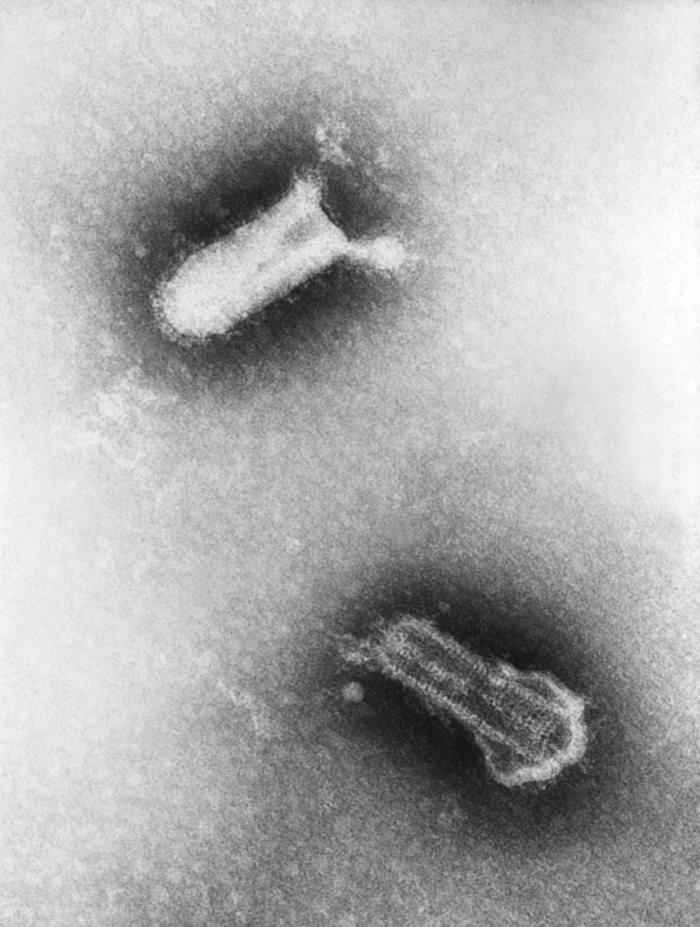
This negatively-stained transmission electron micrograph (TEM) revealed the presence of two Piry virus virions. Note the bullet-like shape of the small 155nm x 162nm virions. Normally, under electron microscopic examination, the virions are observed as being discoidal or spheroidal in shape, and only rarely as bullet-shaped, as was the case here.Created: 1975
-
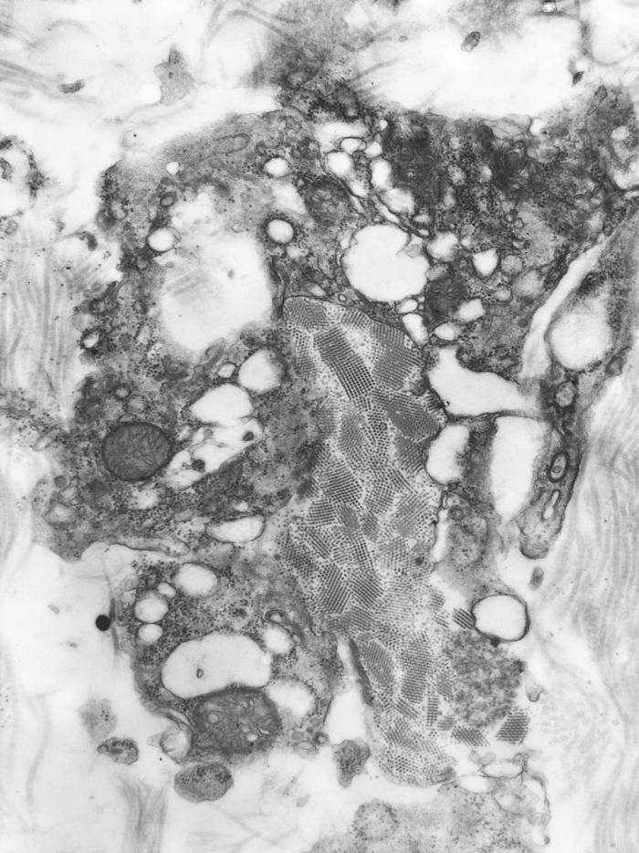
This colorized transmission electron micrograph (TEM) depicts a salivary gland that had been extracted from a mosquito, which was infected by the Eastern equine encephalitis (EEE) virus, which has been colorized red; magnified 83,900x.Created: 1968
-
This negatively-stained transmission electron micrograph (TEM) revealed the presence of numerous Piry virus virions, many of which could be seen as they were budding from the host cell, thereby, becoming free to migrate throughout the hosts system. Note the bullet-like shape of the small 155nm x 162nm virions, as theyre freed from the host cell. Normally, under electron microscopic examination, the virions are observed as being discoidal or spheroidal in shape, and only rarely as bullet-shaped, as was the case here.Created: 1975
-
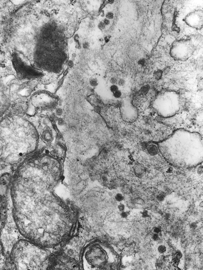
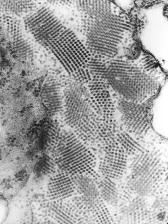
This 1975 transmission electron micrograph (TEM) revealed the presence of a number of Eastern Equine Encephalitis (EEE) virus virions that happened to be in a specimen of central nervous system tissue. EEE is an zoonotic arbovirus, which means that its spead to human beings through the bite of an infected mosquito. EEE virus (EEEV) occurs in the eastern half of the United States where it causes disease in humans, horses, and some bird species. Because of the high mortality rate, EEE is regarded as one of the most serious mosquito-borne diseases in the United States. EEE is a Togaviridae virus family member, and the genus Alphavirus.Created: 1975
-
This negatively-stained transmission electron micrograph (TEM) revealed the presence of numerous Reovirus type-3 virions. This virus organism is a member of the family, Reoviridae, genera of which include the Coltivirus, i.e., Colorado tick fever, Orbivirus, i.e., bluetongue virus, and Orthoreovius, within which belong these virions.Created: 1975