-
-
-
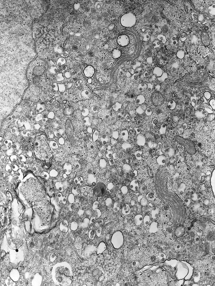
This 1975 transmission electron micrograph (TEM) revealed the presence of a number of infectious bronchitis virus (IBV) virions, which are Coronaviridae family members, and members of the genus Coronavirus. IBV is a highly contagious pathogen, which infects poultry of all ages, affecting a number of organ systems including the respiratory and urogenital organs. IBV is categorized as a Group 3 coronavirus member, with a helical genome composed of positive-sense single-stranded RNA ((+) ssRNA). This is an enveloped virus, which means that its outermost covering is derived from the host cell membrane.Created: 1975
-
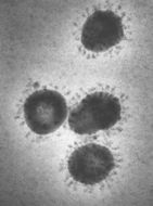
This negatively-stained 1975 transmission electron micrograph (TEM) revealed the presence of a number of human cooronavirus, HCoV-229E virions. This organism is a member of the family, Coronaviridae, and the genus Coronavirus. The coronavirus helical genome is composed of positive-sense single-stranded RNA ((+) ssRNA). This is an enveloped virus, which means that its outermost covering is derived from the host cell membrane.Created: 1975
-
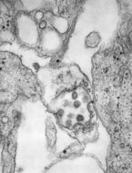
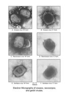
This montage of six transmission electron micrographs (TEM) depicted three different Poxviridae family members, and their morphologic similarities in both their M, or mulberry form, and their C, or capsular form. The three viruses include the cowpox, raccoonpox, and the gerbilpox viruses.The Poxviridae viruses carry at their core, a genome composed of a single, linear double-stranded DNA segment. Before its eradication, one of the most infamous viruses known, the smallpox virus, Variola major, was a member of this family.Created: 1975
-
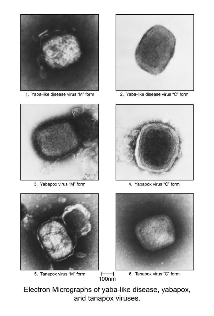
This compilation of a number of transmission electron micrographs (TEM), revealed some of the morphologic differences displayed by three Poxviridae family members: Yaba-like disease (YLD) virus, Yabapox virus, and Tanapox virus, each in its M and C forms. M form represents the virions in their mulberry configuration, whereupon, the capsid appears much like a mulberry, and the C form represents the capsular appearance of the virions capsid, or external protein coat.Created:
-
Highly magnified at 310,000X, this negative-stained transmission electron micrograph (TEM) depicted a smallpox (variola) virus particle, or a single virion. Variola is a double-stranded DNA virus in the genus, Orthopoxvirus. The virus enters the body via the oropharynx, or respiratory mucosa, spreads systemically, and eventually localizes in small blood vessels of the dermis, which is the layer of skin located below the more superficial epidermis.Created: 1968
-
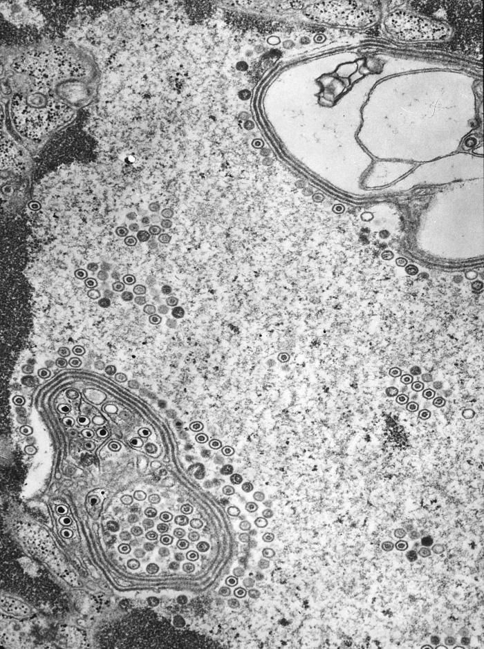
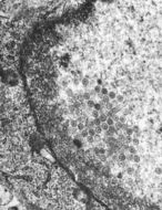
This negatively-stained transmission electron micrograph (TEM) revealed the presence of numerous herpes simplex virions, located inside a cell nucleus in this tissue sample. As members of the Herpesviridae virus family, there are two strains of the herpes simplex virus, HSV-1, which is responsible for cold sores, and HSV-2, which is responsible for genital herpes. At the core of its icosahedral proteinaceous capsid, the HSV contains a double-stranded DNA linear genome.Created: 1975
-
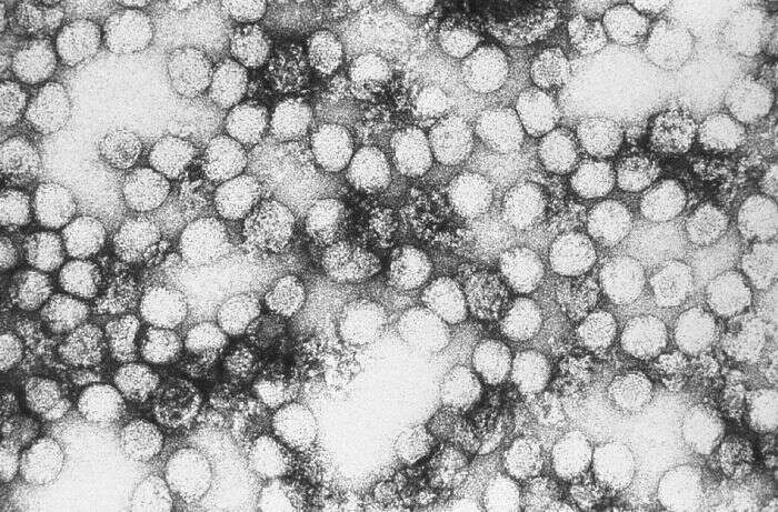
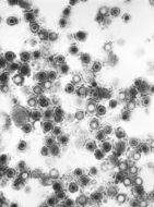
This negatively-stained transmission electron micrograph (TEM) revealed the presence of numerous coronavirus virions in this tissue sample. Coronavirus is a member of the virus family, Coronaviridae, which at their core contain a positive-sense single-stranded RNA genome ((+) ssRNA). Under electronmicrographic examination, the envelope surrounding each virion is studded with a corona of points, hence the derivation of its name, which are actually proteinaceous in nature, and are outcroppings of its envelope's molecular structure.Coronaviruses infect mammals and birds with upper respiratory and gastrointestinal illnesses. The most well-known member of this virus family is the human coronavirus responsible for causing severe acute respiratory syndrome (SARS).Created: 1975
-
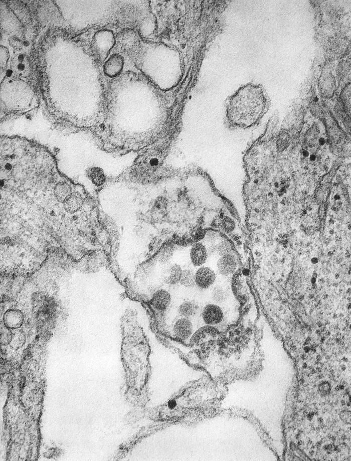
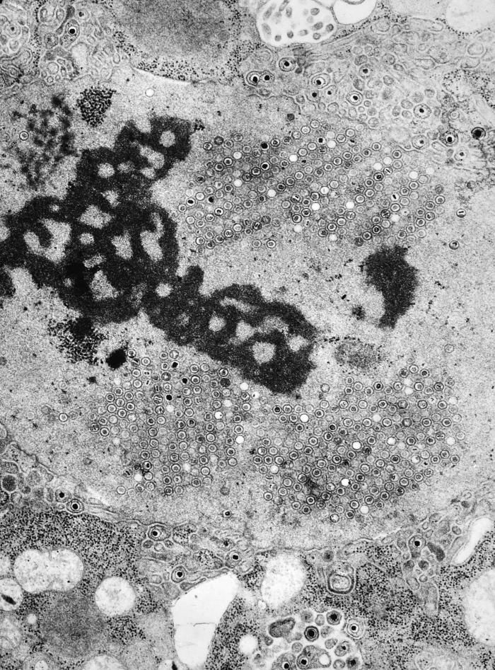
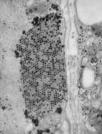
This transmission electron micrograph (TEM) revealed the presence of coxsackie B3 virus particles, which were found within a specimen of muscle tissue. coxsackie B3 virus is a member of the Picornaviridae family of viruses, and the genus, Enterovirus, as is the well-known, nearly eliminated, Poliovirus.Created: 1975
-
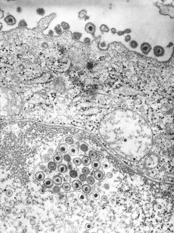
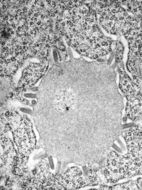
This negatively-stained transmission electron micrograph (TEM) revealed the presence of numerous herpes simplex virions, located both inside the nucleus, and extracellularly in this tissue sample. As members of the Herpesviridae virus family, there are two strains of the herpes simplex virus, HSV-1, which is responsible for cold sores, and HSV-2, which is responsible for genital herpes. At the core of its icosahedral proteinaceous capsid, the HSV contains a double-stranded DNA linear genome.Created: 1975
-
This negatively-stained transmission electron micrograph (TEM) revealed the presence of a number of influenza virus virions. This virus is a Orthomyxoviridae virus family member.Created: 1975
-
This transmission electron micrograph (TEM) revealed the presence of coxsackie B3 virus particles, which were found withing a specimen of muscle tissue. The coxsackie B3 virus is a member of the Picornaviridae family of viruses, and the genus, Enterovirus, as is the well-known, nearly eliminated, Poliovirus.Created: 1975
-
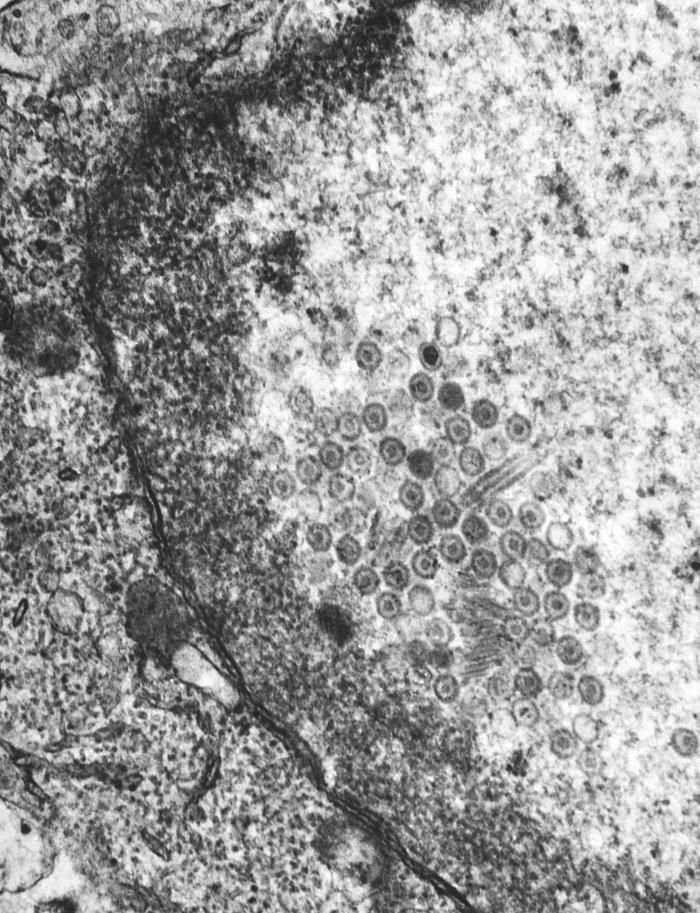
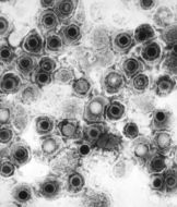
This negatively-stained transmission electron micrograph (TEM) revealed the presence of numerous herpes simplex type-2 virions, members of the Herpesviridae virus family. There are two strains of the herpes simplex virus, HSV-1, which is responsible for cold sores, and HSV-2, which is responsible for genital herpes. At the core of its icosahedral proteinaceous capsid, the HSV contains a double-stranded DNA linear genome.Created: 1975
-
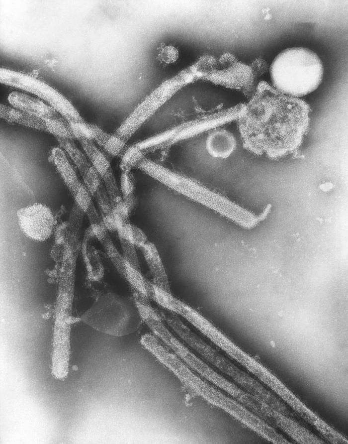
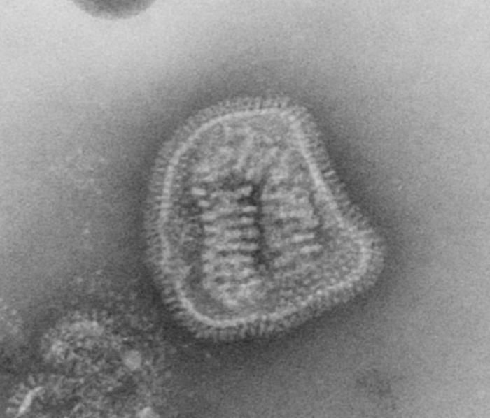
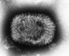
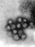
This negatively-stained transmission electron micrograph (TEM) revealed the presence of a number of Hong Kong flu virus virions, the H3N2 subtype of the influenza A virus. This virus is a Orthomyxoviridae virus family member, and was responsible for the flu pandemic of 1968-1969, which infected an estimated 50,000,000 people in the United States, killing 33,000. Note the proteinaceous coat, or capsid, surrounding each virion, and the hemagglutinin-neuraminidase spikes, which differ in terms of their molecular make-up from strain to strain.Created: 1975
-
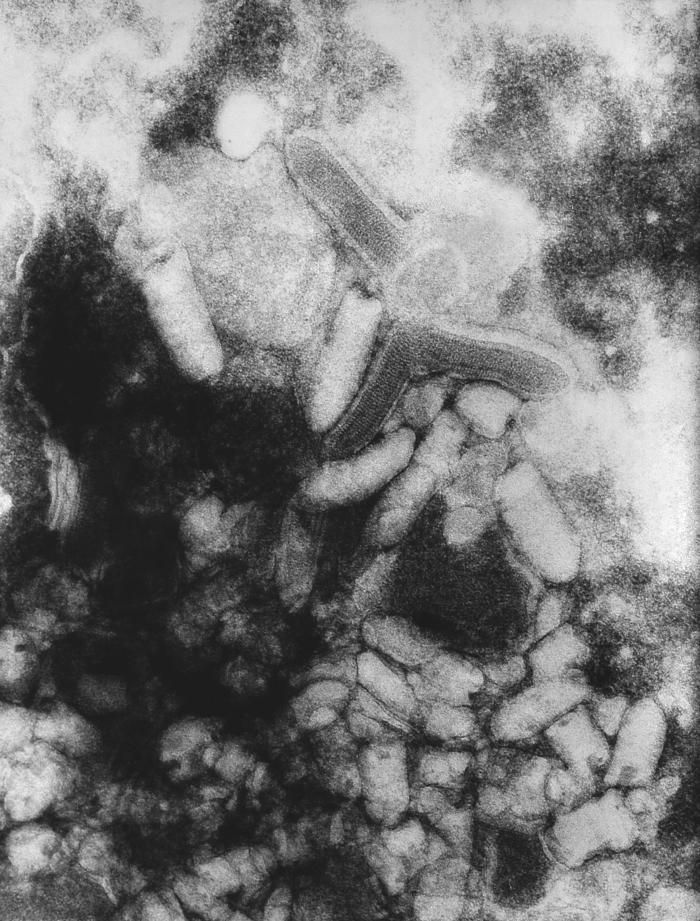
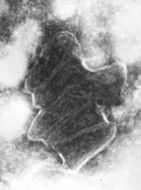
This transmission electron micrograph (TEM) revealed the presence of Lagos bat virus (LBV) virions, and an intracytoplasmic inclusion body in this tissue sample. LBV is a Rhabdoviridae family member, and a member of the genus, Lyssavirus.Created: 1975
-
This negatively-stained transmission electron micrograph (TEM) revealed the presence of numerous herpes simplex virions, members of the Herpesviridae virus family. There are two strains of the herpes simplex virus, HSV-1, which is responsible for cold sores, and HSV-2, which is responsible for genital herpes. At the core of its icosahedral proteinaceous capsid, the HSV contains a double-stranded DNA linear genome.Created: 1975
-
This negatively-stained transmission electron micrograph (TEM) revealed the presence of a number of Hong Kong flu virus virions, the H3N2 subtype of the influenza A virus. This virus is a Orthomyxoviridae virus family member, and was responsible for the flu pandemic of 1968-1969, which infected an estimated 50,000,000 people in the United States, killing 33,000. Note the proteinaceous coat, or capsid, surroundind each virion, and the hemagglutinin-neuraminidase spikes, which differ in terms of their molecular make-up from strain to strain.Created: 1975
-
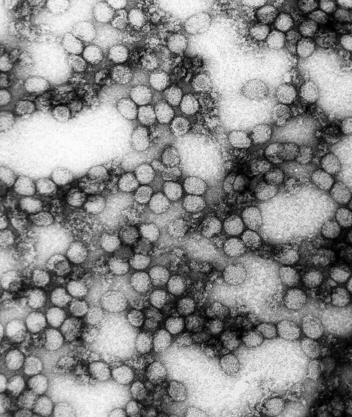
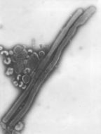
This negatively-stained transmission electron micrograph (TEM) revealed the presence of numerous negative-sense, single-stranded RNA ((-) ssRNA) Flanders virus virions. Note the bullet-like shape of these virions, which are very similar to other Rhabdoviruses, i.e., see PHIL 1876 depicting a TEM revealing the bullet-shaped rabies virus virions.Created: 1975
-
This negatively-stained transmission electron micrograph (TEM) revealed the presence of numerous herpes simplex virions, members of the Herpesviridae virus family. There are two strains of the herpes simplex virus, HSV-1, which is responsible for cold sores, and HSV-2, which is responsible for genital herpes. At the core of its icosahedral proteinaceous capsid, the HSV contains a double-stranded DNA linear genome.Created: 1975
-
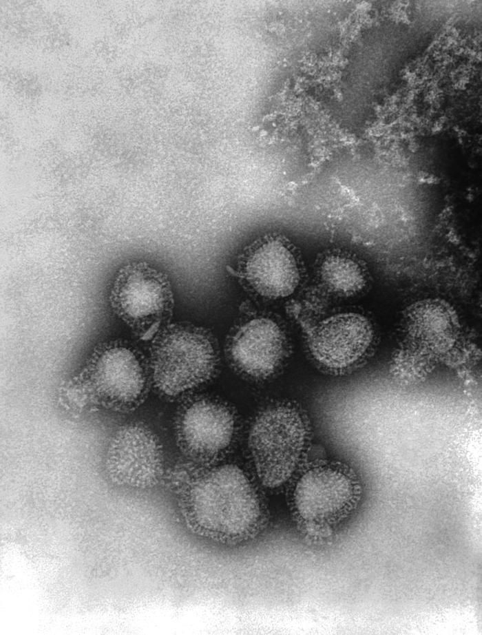
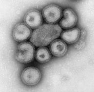
This negative-stained transmission electron micrograph (TEM) depicts the ultrastructural details of a number of influenza virus particles, or virions. A member of the taxonomic family Orthomyxoviridae, the influenza virus is a single-stranded RNA organismThe flu is a contagious respiratory illness caused by influenza viruses. It can cause mild to severe illness, and at times can lead to death. The best way to prevent this illness is by getting a flu vaccination each fall.Every year in the United States, on average:- 5% to 20% of the population gets the flu- more than 200,000 people are hospitalized from flu complications, and- about 36,000 people die from flu. Some people, such as older people, young children, and people with certain health conditions, are at high risk for serious flu complications. See PHIL 10072 for a colorized version of this image.Created: 1973
-
This negatively-stained transmission electron micrograph (TEM) revealed the presence of numerous bovine ephemeral fever virus virions, which are members of the Rhabdoviridae family of viruses, and the genus Ephemerovirus, infecting animals as well as plants.Created: 1975
-
This negatively-stained transmission electron micrograph (TEM) revealed the presence of numerous herpes simplex virions, members of the Herpesviridae virus family. There are two strains of the herpes simplex virus, HSV-1, which is responsible for cold sores, and HSV-2, which is responsible for genital herpes. At the core of its icosahedral proteinaceous capsid, the HSV contains a double-stranded DNA linear genome.Created: 1975
-
This negative-stained transmission electron micrograph (TEM) depicts the ultrastructural details of an influenza virus particle, or virion. A member of the taxonomic family Orthomyxoviridae, the influenza virus is a single-stranded RNA organismThe flu is a contagious respiratory illness caused by influenza viruses. It can cause mild to severe illness, and at times can lead to death. The best way to prevent this illness is by getting a flu vaccination each fall.Every year in the United States, on average:- 5% to 20% of the population gets the flu- more than 200,000 people are hospitalized from flu complications, and- about 36,000 people die from flu. Some people, such as older people, young children, and people with certain health conditions, are at high risk for serious flu complications. See PHIL 10073 for a colorized version of this image.Created: 1981